 Movie
Movie Controller

[English] 日本語
 Yorodumi
- PDB-1em8: Crystal structure of chi and psi subunit heterodimer from DNA POL III -
+ Open data

- Basic information
| Entry | Database: PDB / ID: 1em8 | ||||||
|---|---|---|---|---|---|---|---|
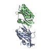
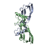
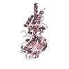
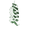
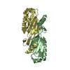
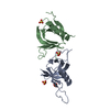
| Title | Crystal structure of chi and psi subunit heterodimer from DNA POL III | ||||||
 Components Components |
| ||||||
 Keywords Keywords | GENE REGULATION / DNA Pol III / heterodimer / clamp-loader / alpha-beta fold | ||||||
| Function / homology |  Function and homology information Function and homology informationDNA polymerase III, clamp loader complex / DNA polymerase III complex / replisome / positive regulation of DNA-templated DNA replication initiation / 3'-5' exonuclease activity / response to radiation / DNA-templated DNA replication / DNA-directed DNA polymerase / DNA-directed DNA polymerase activity / DNA replication / DNA binding Similarity search - Function | ||||||
| Biological species |  | ||||||
| Method |  X-RAY DIFFRACTION / Resolution: 2.1 Å X-RAY DIFFRACTION / Resolution: 2.1 Å | ||||||
 Authors Authors | Gulbis, J.M. / Finkelstein, J. / O'Donnell, M. / Kuriyan, J. | ||||||
 Citation Citation | Journal: Eur.J.Biochem. / Year: 2004 Title: Crystal structure of the chi:psi sub-assembly of the Escherichia coli DNA polymerase clamp-loader complex. Authors: Gulbis, J.M. / Kazmirski, S.L. / Finkelstein, J. / Kelman, Z. / O'Donnell, M. / Kuriyan, J. | ||||||
| History |
|
- Structure visualization
| Structure viewer | Molecule: MolmilJmol/JSmol |
|---|
- Downloads & links
-Download
| PDBx/mmCIF format | 1em8.cif.gz | 112.6 KB | Display | PDBx/mmCIF format |
|---|---|---|---|---|
| PDB format | pdb1em8.ent.gz | 87.7 KB | Display | PDB format |
| PDBx/mmJSON format | 1em8.json.gz | Tree view | PDBx/mmJSON format | |
| Others |  Other downloads Other downloads |
-Validation report
| Arichive directory | https://data.pdbj.org/pub/pdb/validation_reports/em/1em8ftp://data.pdbj.org/pub/pdb/validation_reports/em/1em8 | HTTPS FTP |
|---|
-Related structure data
| Similar structure data |
|---|

-Links
 PDBj
PDBj
- Assembly
| Deposited unit | 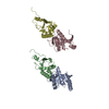
| ||||||||
|---|---|---|---|---|---|---|---|---|---|
| 1 | 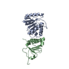
| ||||||||
| 2 | 
| ||||||||
| Unit cell |
| ||||||||
| Details | One biological assembly is a heterodimer constructed from chain A and chain B / One biological assembly is a heterodimer constructed from chain C and chain D |
-Components
| #1: Protein | Mass: 16653.742 Da / Num. of mol.: 2 / Fragment: RESIDUES 1-147 Source method: isolated from a genetically manipulated source Source: (gene. exp.) #2: Protein | Mass: 12203.835 Da / Num. of mol.: 2 / Fragment: RESIDUES 26-137 Source method: isolated from a genetically manipulated source Source: (gene. exp.) #3: Water | ChemComp-HOH / |  Mass: 18.015 Da / Num. of mol.: 256 / Fragment: WATER / Source method: isolated from a natural source / Formula: H2O Mass: 18.015 Da / Num. of mol.: 256 / Fragment: WATER / Source method: isolated from a natural source / Formula: H2O |
|---|
-Experimental details
-Experiment
| Experiment | Method: X-RAY DIFFRACTION / Number of used crystals: 1 |
|---|
- Sample preparation
| Crystal | Density Matthews: 2.42 Å3/Da / Density % sol: 49.09 % | |||||||||||||||||||||||||||||||||||||||||||||||||
|---|---|---|---|---|---|---|---|---|---|---|---|---|---|---|---|---|---|---|---|---|---|---|---|---|---|---|---|---|---|---|---|---|---|---|---|---|---|---|---|---|---|---|---|---|---|---|---|---|---|---|
| Crystal grow | Temperature: 277 K / Method: vapor diffusion, hanging drop / pH: 6.8 Details: methylpentanediol, HEPES, PEG 4000, Glycerol, pH 6.8, VAPOR DIFFUSION, HANGING DROP, temperature 277K | |||||||||||||||||||||||||||||||||||||||||||||||||
| Crystal grow | *PLUS Temperature: 4 K / pH: 7.5 / Method: vapor diffusion, hanging drop | |||||||||||||||||||||||||||||||||||||||||||||||||
| Components of the solutions | *PLUS
|
-Data collection
| Diffraction | Mean temperature: 100 K |
|---|---|
| Diffraction source | Source: ROTATING ANODE / Type: RIGAKU / Wavelength: 1.5418 |
| Detector | Type: RIGAKU / Detector: IMAGE PLATE / Date: Jan 1, 1994 |
| Radiation | Protocol: SINGLE WAVELENGTH / Monochromatic (M) / Laue (L): M / Scattering type: x-ray |
| Radiation wavelength | Wavelength: 1.5418 Å / Relative weight: 1 |
| Reflection | Resolution: 2.1→20 Å / Num. all: 223463 / Num. obs: 218771 / % possible obs: 97.9 % / Observed criterion σ(F): 3 / Observed criterion σ(I): 3 / Redundancy: 6.8 % / Biso Wilson estimate: 24 Å2 / Rmerge(I) obs: 0.066 / Net I/σ(I): 13.2 |
| Reflection shell | Resolution: 2.1→2.2 Å / Redundancy: 1 % / Rmerge(I) obs: 0.194 / Num. unique all: 4047 / % possible all: 95.2 |
| Reflection | *PLUS Lowest resolution: 20 Å / Num. obs: 32326 / Num. measured all: 218771 |
| Reflection shell | *PLUS % possible obs: 95.2 % |
- Processing
| Software |
| |||||||||||||||||||||||||
|---|---|---|---|---|---|---|---|---|---|---|---|---|---|---|---|---|---|---|---|---|---|---|---|---|---|---|
| Refinement | Resolution: 2.1→20 Å / σ(F): 0 / σ(I): 0 / Stereochemistry target values: Engh & Huber / Details: maximum likelihood
| |||||||||||||||||||||||||
| Refinement step | Cycle: LAST / Resolution: 2.1→20 Å
| |||||||||||||||||||||||||
| Refine LS restraints |
| |||||||||||||||||||||||||
| Refinement | *PLUS Lowest resolution: 20 Å | |||||||||||||||||||||||||
| Solvent computation | *PLUS | |||||||||||||||||||||||||
| Displacement parameters | *PLUS | |||||||||||||||||||||||||
| Refine LS restraints | *PLUS
|
