Redundancy: 6.3 % / Av σ(I) over netI: 33.54 / Number: 581055 / Rmerge(I) obs: 0.102 / Χ2: 1.02 / D res high: 2.01 Å / D res low: 50 Å / Num. obs: 92883 / % possible obs: 99.6
Diffraction reflection shell
Highest resolution (Å)
Lowest resolution (Å)
% possible obs (%)
ID
Rmerge(I) obs
Chi squared
Redundancy
4.33
50
98.9
1
0.051
1.025
6.5
3.44
4.33
99.8
1
0.057
1.007
6.5
3
3.44
99.9
1
0.076
1.001
6.5
2.73
3
100
1
0.111
1.027
6.4
2.53
2.73
99.9
1
0.155
1.027
6.3
2.38
2.53
99.9
1
0.195
1.032
6.2
2.26
2.38
99.8
1
0.24
1.033
6.2
2.17
2.26
99.5
1
0.322
1.16
6.1
2.08
2.17
99.2
1
0.376
1.002
6.1
2.01
2.08
99.3
1
0.445
0.913
5.9
Reflection
Resolution: 1.76→50 Å / Num. obs: 72469 / % possible obs: 99 % / Redundancy: 7.2 % / Rmerge(I) obs: 0.054 / Χ2: 1.004 / Net I/σ(I): 18.7
Reflection shell
Resolution (Å)
Redundancy (%)
Rmerge(I) obs
Num. unique all
Χ2
% possible all
1.76-1.82
5.6
0.256
6901
0.847
95.7
1.82-1.9
5.9
0.179
7243
0.885
99.9
1.9-1.98
5.9
0.128
7220
0.978
100
1.98-2.09
6
0.092
7239
1.028
100
2.09-2.22
6
0.069
7269
1.014
100
2.22-2.39
6
0.053
7241
1.026
100
2.39-2.63
6
0.044
7307
1.083
100
2.63-3.01
6.2
0.043
7331
1.04
99.9
3.01-3.79
12.1
0.061
7335
1.065
99.1
3.79-50
11.6
0.046
7383
0.99
96.1
-
Processing
Software
Name
Version
Classification
NB
DENZO
datareduction
SCALEPACK
datascaling
REFMAC
5.5.0066
refinement
PDB_EXTRACT
3.005
dataextraction
Refinement
Method to determine structure: MAD / Resolution: 1.76→37.807 Å / Cor.coef. Fo:Fc: 0.954 / Cor.coef. Fo:Fc free: 0.938 / Occupancy max: 1 / Occupancy min: 0.5 / SU B: 1.961 / SU ML: 0.065 / Cross valid method: THROUGHOUT / σ(F): 0 / ESU R: 0.107 / ESU R Free: 0.109 / Stereochemistry target values: MAXIMUM LIKELIHOOD Details: HYDROGENS HAVE BEEN ADDED IN THE RIDING POSITIONS. U VALUES: REFINED INDIVIDUALLY
Rfactor
Num. reflection
% reflection
Selection details
Rfree
0.222
3641
5 %
RANDOM
Rwork
0.182
-
-
-
obs
0.184
72414
98.94 %
-
Solvent computation
Ion probe radii: 0.8 Å / Shrinkage radii: 0.8 Å / VDW probe radii: 1.4 Å / Solvent model: MASK
In the structure databanks used in Yorodumi, some data are registered as the other names, "COVID-19 virus" and "2019-nCoV". Here are the details of the virus and the list of structure data.
Jan 31, 2019. EMDB accession codes are about to change! (news from PDBe EMDB page)
EMDB accession codes are about to change! (news from PDBe EMDB page)
The allocation of 4 digits for EMDB accession codes will soon come to an end. Whilst these codes will remain in use, new EMDB accession codes will include an additional digit and will expand incrementally as the available range of codes is exhausted. The current 4-digit format prefixed with “EMD-” (i.e. EMD-XXXX) will advance to a 5-digit format (i.e. EMD-XXXXX), and so on. It is currently estimated that the 4-digit codes will be depleted around Spring 2019, at which point the 5-digit format will come into force.
The EM Navigator/Yorodumi systems omit the EMD- prefix.
Related info.:Q: What is EMD? / ID/Accession-code notation in Yorodumi/EM Navigator
Yorodumi is a browser for structure data from EMDB, PDB, SASBDB, etc.
This page is also the successor to EM Navigator detail page, and also detail information page/front-end page for Omokage search.
The word "yorodu" (or yorozu) is an old Japanese word meaning "ten thousand". "mi" (miru) is to see.
Related info.:EMDB / PDB / SASBDB / Comparison of 3 databanks / Yorodumi Search / Aug 31, 2016. New EM Navigator & Yorodumi / Yorodumi Papers / Jmol/JSmol / Function and homology information / Changes in new EM Navigator and Yorodumi
 Movie
Movie Controller

 Yorodumi
 Open data
 Basic information
 Components
 Keywords
 Function and homology information
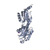
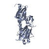
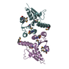
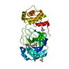
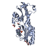
 Vibrio parahaemolyticus (bacteria)
 X-RAY DIFFRACTION /
  Authors
 Citation
 Structure visualization
 Downloads & links
 Other downloads


 PDBj
PDBj


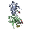
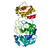
 Assembly
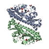

 Mass: 94.971 Da / Num. of mol.: 2 / Source method: obtained synthetically / Formula: PO4
 Mass: 35.453 Da / Num. of mol.: 2 / Source method: obtained synthetically / Formula: Cl
 Mass: 62.068 Da / Num. of mol.: 9 / Source method: obtained synthetically / Formula: C2H6O2
 Mass: 18.015 Da / Num. of mol.: 584 / Source method: isolated from a natural source / Formula: H2O
 Sample preparation
 Processing