[English] 日本語
 Yorodumi
- PDB-3jqg: Crystal structure of pteridine reductase 1 (PTR1) from Trypanosom... -
+ Open data
- Basic information
| Entry | Database: PDB / ID: 3jqg | |||||||||
|---|---|---|---|---|---|---|---|---|---|---|
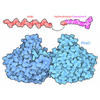
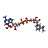
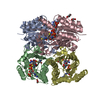
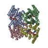
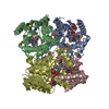
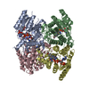
| Title | Crystal structure of pteridine reductase 1 (PTR1) from Trypanosoma brucei in ternary complex with cofactor (NADP+) and inhibitor 6-[(4-methoxybenzyl)sulfanyl]pyrimidine-2,4-diamine (AX6) | |||||||||
 Components Components | (Pteridine reductase 1) x 2 | |||||||||
 Keywords Keywords | OXIDOREDUCTASE / PTERIDINE REDUCTASE / PTR1 / TRYPANOSOMA BRUCEI / SHORT CHAIN DEHYDROGENASE / INHIBITOR | |||||||||
| Function / homology |  Function and homology information Function and homology informationpteridine reductase / pteridine reductase activity / oxidoreductase activity / nucleotide binding / cytosol Similarity search - Function | |||||||||
| Biological species |  | |||||||||
| Method |  X-RAY DIFFRACTION / X-RAY DIFFRACTION /  SYNCHROTRON / SYNCHROTRON /  MOLECULAR REPLACEMENT / MOLECULAR REPLACEMENT /  molecular replacement / Resolution: 1.9 Å molecular replacement / Resolution: 1.9 Å | |||||||||
 Authors Authors | Tulloch, L.B. / Hunter, W.N. | |||||||||
 Citation Citation |  Journal: J.Med.Chem. / Year: 2010 Journal: J.Med.Chem. / Year: 2010Title: Structure-based design of pteridine reductase inhibitors targeting african sleeping sickness and the leishmaniases. Authors: Tulloch, L.B. / Martini, V.P. / Iulek, J. / Huggan, J.K. / Lee, J.H. / Gibson, C.L. / Smith, T.K. / Suckling, C.J. / Hunter, W.N. | |||||||||
| History |
|
- Structure visualization
| Structure viewer | Molecule:  Molmil Molmil Jmol/JSmol Jmol/JSmol |
|---|
- Downloads & links
- Download
| PDBx/mmCIF format |  3jqg.cif.gz 3jqg.cif.gz | 218.4 KB | Display |  PDBx/mmCIF format PDBx/mmCIF format |
|---|---|---|---|---|
| PDB format |  pdb3jqg.ent.gz pdb3jqg.ent.gz | 172.8 KB | Display |  PDB format PDB format |
| PDBx/mmJSON format |  3jqg.json.gz 3jqg.json.gz | Tree view |  PDBx/mmJSON format PDBx/mmJSON format | |
| Others |  Other downloads Other downloads |
-Validation report
| Summary document |  3jqg_validation.pdf.gz 3jqg_validation.pdf.gz | 2.9 MB | Display |  wwPDB validaton report wwPDB validaton report |
|---|---|---|---|---|
| Full document |  3jqg_full_validation.pdf.gz 3jqg_full_validation.pdf.gz | 2.8 MB | Display | |
| Data in XML |  3jqg_validation.xml.gz 3jqg_validation.xml.gz | 45.7 KB | Display | |
| Data in CIF |  3jqg_validation.cif.gz 3jqg_validation.cif.gz | 63.9 KB | Display | |
| Arichive directory |  https://data.pdbj.org/pub/pdb/validation_reports/jq/3jqg https://data.pdbj.org/pub/pdb/validation_reports/jq/3jqg ftp://data.pdbj.org/pub/pdb/validation_reports/jq/3jqg ftp://data.pdbj.org/pub/pdb/validation_reports/jq/3jqg | HTTPS FTP |
-Related structure data
| Related structure data | 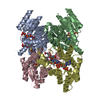 3bmcC  3bmnC  3bmoC  3bmqC  3jq6C  3jq7C  3jq8C  3jq9C  3jqaC  3jqbC  3jqcC  3jqdC  3jqeC  3jqfC 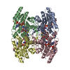 2c7vS C: citing same article ( S: Starting model for refinement |
|---|---|
| Similar structure data |
- Links
Links
- Assembly
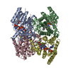
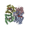
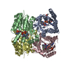
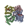
| Deposited unit | 
| |||||||||||||||||||||||||||||||||||||||||||||
|---|---|---|---|---|---|---|---|---|---|---|---|---|---|---|---|---|---|---|---|---|---|---|---|---|---|---|---|---|---|---|---|---|---|---|---|---|---|---|---|---|---|---|---|---|---|---|
| 1 |
| |||||||||||||||||||||||||||||||||||||||||||||
| Unit cell |
| |||||||||||||||||||||||||||||||||||||||||||||
| Noncrystallographic symmetry (NCS) | NCS domain:
NCS domain segments:
|
- Components
| #1: Protein | Mass: 30685.787 Da / Num. of mol.: 2 / Fragment: UNP residues 102-369 Source method: isolated from a genetically manipulated source Source: (gene. exp.)   #2: Protein | Mass: 30669.791 Da / Num. of mol.: 2 / Fragment: UNP residues 102-369 Source method: isolated from a genetically manipulated source Source: (gene. exp.)   #3: Chemical | ChemComp-NAP / #4: Chemical | ChemComp-AX6 / #5: Water | ChemComp-HOH / | |
|---|
-Experimental details
-Experiment
| Experiment | Method:  X-RAY DIFFRACTION / Number of used crystals: 1 X-RAY DIFFRACTION / Number of used crystals: 1 |
|---|
- Sample preparation
| Crystal | Density Matthews: 2.03 Å3/Da / Density % sol: 39.4 % |
|---|---|
| Crystal grow | Temperature: 293 K / Method: vapor diffusion Details: 2-3M Sodium acetate, 10-100mM Sodium citrate, pH 4.0-6.0, VAPOR DIFFUSION, temperature 293K PH range: 4.0-6.0 |
-Data collection
| Diffraction | Mean temperature: 100 K |
|---|---|
| Diffraction source | Source:  SYNCHROTRON / Site: SYNCHROTRON / Site:  ESRF ESRF  / Beamline: ID23-2 / Wavelength: 0.873 Å / Beamline: ID23-2 / Wavelength: 0.873 Å |
| Detector | Type: MARMOSAIC 225 mm CCD / Detector: CCD / Date: May 20, 2006 Details: Pt coated mirrors in a Kirkpatrick-Baez (KB) geometry |
| Radiation | Monochromator: Si(111) / Protocol: SINGLE WAVELENGTH / Monochromatic (M) / Laue (L): M / Scattering type: x-ray |
| Radiation wavelength | Wavelength: 0.873 Å / Relative weight: 1 |
| Reflection | Resolution: 1.9→53.2 Å / Num. obs: 74417 / % possible obs: 96.5 % / Redundancy: 2.4 % / Rmerge(I) obs: 0.092 |
| Reflection shell | Resolution: 1.9→2.01 Å / Redundancy: 2.7 % / Rmerge(I) obs: 0.29 / Mean I/σ(I) obs: 1.9 |
-Phasing
| Phasing | Method:  molecular replacement molecular replacement |
|---|
- Processing
| Software |
| |||||||||||||||||||||||||||||||||||||||||||||||||||||||||||||||||||||||||||||||||||||||||||||||||||||||||||||||||||||||||||||
|---|---|---|---|---|---|---|---|---|---|---|---|---|---|---|---|---|---|---|---|---|---|---|---|---|---|---|---|---|---|---|---|---|---|---|---|---|---|---|---|---|---|---|---|---|---|---|---|---|---|---|---|---|---|---|---|---|---|---|---|---|---|---|---|---|---|---|---|---|---|---|---|---|---|---|---|---|---|---|---|---|---|---|---|---|---|---|---|---|---|---|---|---|---|---|---|---|---|---|---|---|---|---|---|---|---|---|---|---|---|---|---|---|---|---|---|---|---|---|---|---|---|---|---|---|---|---|
| Refinement | Method to determine structure:  MOLECULAR REPLACEMENT MOLECULAR REPLACEMENTStarting model: PDB entry 2C7V Resolution: 1.9→53.2 Å / Cor.coef. Fo:Fc: 0.94 / Cor.coef. Fo:Fc free: 0.905 / WRfactor Rfree: 0.272 / WRfactor Rwork: 0.218 / Occupancy max: 1 / Occupancy min: 0.1 / FOM work R set: 0.807 / SU B: 9.514 / SU ML: 0.141 / SU R Cruickshank DPI: 0.195 / SU Rfree: 0.179 / TLS residual ADP flag: LIKELY RESIDUAL / Cross valid method: THROUGHOUT / σ(F): 0 / ESU R: 0.195 / ESU R Free: 0.179 / Stereochemistry target values: MAXIMUM LIKELIHOOD
| |||||||||||||||||||||||||||||||||||||||||||||||||||||||||||||||||||||||||||||||||||||||||||||||||||||||||||||||||||||||||||||
| Solvent computation | Ion probe radii: 0.8 Å / Shrinkage radii: 0.8 Å / VDW probe radii: 1.4 Å / Solvent model: MASK | |||||||||||||||||||||||||||||||||||||||||||||||||||||||||||||||||||||||||||||||||||||||||||||||||||||||||||||||||||||||||||||
| Displacement parameters | Biso max: 70.24 Å2 / Biso mean: 17.855 Å2 / Biso min: 2 Å2
| |||||||||||||||||||||||||||||||||||||||||||||||||||||||||||||||||||||||||||||||||||||||||||||||||||||||||||||||||||||||||||||
| Refinement step | Cycle: LAST / Resolution: 1.9→53.2 Å
| |||||||||||||||||||||||||||||||||||||||||||||||||||||||||||||||||||||||||||||||||||||||||||||||||||||||||||||||||||||||||||||
| Refine LS restraints |
| |||||||||||||||||||||||||||||||||||||||||||||||||||||||||||||||||||||||||||||||||||||||||||||||||||||||||||||||||||||||||||||
| Refine LS restraints NCS | Dom-ID: 1 / Ens-ID: 1 / Number: 1797 / Refine-ID: X-RAY DIFFRACTION
| |||||||||||||||||||||||||||||||||||||||||||||||||||||||||||||||||||||||||||||||||||||||||||||||||||||||||||||||||||||||||||||
| LS refinement shell | Resolution: 1.9→1.949 Å / Total num. of bins used: 20
| |||||||||||||||||||||||||||||||||||||||||||||||||||||||||||||||||||||||||||||||||||||||||||||||||||||||||||||||||||||||||||||
| Refinement TLS params. | Method: refined / Refine-ID: X-RAY DIFFRACTION
| |||||||||||||||||||||||||||||||||||||||||||||||||||||||||||||||||||||||||||||||||||||||||||||||||||||||||||||||||||||||||||||
| Refinement TLS group |
|
 Movie
Movie Controller












 PDBj
PDBj


