 Movie
Movie Controller

[English] 日本語
 Yorodumi
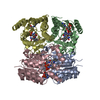
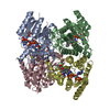
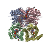
- PDB-2c7v: Structure of Trypanosoma brucei pteridine reductase (PTR1) in ter... -
+ Open data

- Basic information
| Entry | Database: PDB / ID: 2c7v | ||||||
|---|---|---|---|---|---|---|---|
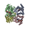
| Title | Structure of Trypanosoma brucei pteridine reductase (PTR1) in ternary complex with cofactor and the antifolate methotrexate | ||||||
 Components Components | PTERIDINE REDUCTASE | ||||||
 Keywords Keywords | OXIDOREDUCTASE / PTERIDINE REDUCTASE / TRYPANOSOMATIDS / DRUG RESISTANCE / SHORT-CHAIN DEHYDROGENASE/REDUCTASE / METHOTREXATE RESISTANCE | ||||||
| Function / homology |  Function and homology information Function and homology information | ||||||
| Biological species |  | ||||||
| Method |  X-RAY DIFFRACTION / MOLECULAR REPLACEMENT / Resolution: 2.2 Å X-RAY DIFFRACTION / MOLECULAR REPLACEMENT / Resolution: 2.2 Å | ||||||
 Authors Authors | Dawson, A. / Gibellini, F. / Sienkiewicz, N. / Fyfe, P.K. / McLuskey, K. / Fairlamb, A.H. / Hunter, W.N. | ||||||
 Citation Citation | Journal: Mol.Microbiol. / Year: 2006 Title: Structure and Reactivity of Trypanosoma Brucei Pteridine Reductase: Inhibition by the Archetypal Antifolate Methotrexate Authors: Dawson, A. / Gibellini, F. / Sienkiewicz, N. / Tulloch, L.B. / Fyfe, P.K. / Mcluskey, K. / Fairlamb, A.H. / Hunter, W.N. | ||||||
| History |
|
- Structure visualization
| Structure viewer | Molecule: MolmilJmol/JSmol |
|---|
- Downloads & links
-Download
| PDBx/mmCIF format | 2c7v.cif.gz | 228.1 KB | Display | PDBx/mmCIF format |
|---|---|---|---|---|
| PDB format | pdb2c7v.ent.gz | 184.2 KB | Display | PDB format |
| PDBx/mmJSON format | 2c7v.json.gz | Tree view | PDBx/mmJSON format | |
| Others |  Other downloads Other downloads |
-Validation report
| Arichive directory | https://data.pdbj.org/pub/pdb/validation_reports/c7/2c7vftp://data.pdbj.org/pub/pdb/validation_reports/c7/2c7v | HTTPS FTP |
|---|
-Related structure data
| Related structure data | 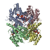 1e92S S: Starting model for refinement |
|---|---|
| Similar structure data |


-Links
 PDBj
PDBj


- Assembly
| Deposited unit | 
| ||||||||||||||||
|---|---|---|---|---|---|---|---|---|---|---|---|---|---|---|---|---|---|
| 1 |
| ||||||||||||||||
| Unit cell |
| ||||||||||||||||
| Noncrystallographic symmetry (NCS) | NCS oper:
|
-Components
-Protein , 1 types, 4 molecules ABCD
| #1: Protein | Mass: 28738.406 Da / Num. of mol.: 4 Source method: isolated from a genetically manipulated source Source: (gene. exp.)  |
|---|
-Non-polymers , 5 types, 811 molecules 
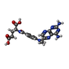


| #2: Chemical | ChemComp-NAP /  Mass: 743.405 Da / Num. of mol.: 4 / Source method: obtained synthetically / Formula: C21H28N7O17P3 Mass: 743.405 Da / Num. of mol.: 4 / Source method: obtained synthetically / Formula: C21H28N7O17P3#3: Chemical | ChemComp-MTX /  Mass: 454.439 Da / Num. of mol.: 4 / Source method: obtained synthetically / Formula: C20H22N8O5 Mass: 454.439 Da / Num. of mol.: 4 / Source method: obtained synthetically / Formula: C20H22N8O5#4: Chemical |  Mass: 58.693 Da / Num. of mol.: 2 / Source method: obtained synthetically / Formula: Ni Mass: 58.693 Da / Num. of mol.: 2 / Source method: obtained synthetically / Formula: Ni#5: Chemical |  Mass: 59.044 Da / Num. of mol.: 2 / Source method: obtained synthetically / Formula: C2H3O2 Mass: 59.044 Da / Num. of mol.: 2 / Source method: obtained synthetically / Formula: C2H3O2#6: Water | ChemComp-HOH / | Mass: 18.015 Da / Num. of mol.: 799 / Source method: isolated from a natural source / Formula: H2O |
|---|
-Details
| Has protein modification | Y |
|---|
-Experimental details
-Experiment
| Experiment | Method: X-RAY DIFFRACTION / Number of used crystals: 1 |
|---|
- Sample preparation
| Crystal | Density Matthews: 2.01 Å3/Da / Density % sol: 38.22 % |
|---|---|
| Crystal grow | Method: vapor diffusion, hanging drop / pH: 7 Details: PROTEIN BUFFER WAS 20 MM TRIS HCL, PH 7.0 WITH 1MM NADP, 1 MM METHOTREXATE AND 20 MM DITHIOTHREITOL, PROTEIN CONCENTRATION WAS 6 MG/ML. HANGING DROP RESERVOIR SOLUTION WAS 0.1 M SODIUM ...Details: PROTEIN BUFFER WAS 20 MM TRIS HCL, PH 7.0 WITH 1MM NADP, 1 MM METHOTREXATE AND 20 MM DITHIOTHREITOL, PROTEIN CONCENTRATION WAS 6 MG/ML. HANGING DROP RESERVOIR SOLUTION WAS 0.1 M SODIUM CACODYLATE PH 6.5 AND 1.4 M SODIUM ACETATE. |
-Data collection
| Diffraction | Mean temperature: 100 K |
|---|---|
| Diffraction source | Source: ROTATING ANODE / Type: RIGAKU MICROMAX-007 / Wavelength: 1.5418 |
| Detector | Type: RIGAKU IMAGE PLATE / Detector: IMAGE PLATE / Date: Jul 30, 2004 / Details: MIRRORS |
| Radiation | Protocol: SINGLE WAVELENGTH / Monochromatic (M) / Laue (L): M / Scattering type: x-ray |
| Radiation wavelength | Wavelength: 1.5418 Å / Relative weight: 1 |
| Reflection | Resolution: 2.2→74.5 Å / Num. obs: 50048 / % possible obs: 99.2 % / Redundancy: 2.8 % / Rmerge(I) obs: 0.06 / Net I/σ(I): 12.4 |
| Reflection shell | Resolution: 2.2→2.26 Å / Redundancy: 2.1 % / Rmerge(I) obs: 0.17 / Mean I/σ(I) obs: 3.5 / % possible all: 93.1 |
- Processing
| Software |
| ||||||||||||||||||||||||||||||||||||||||||||||||||||||||||||||||||||||||||||||||||||||||||||||||||||||||||||||||||||||||||||||||||||||||||||||||||||||||||||||||||||||||||||||||||||||
|---|---|---|---|---|---|---|---|---|---|---|---|---|---|---|---|---|---|---|---|---|---|---|---|---|---|---|---|---|---|---|---|---|---|---|---|---|---|---|---|---|---|---|---|---|---|---|---|---|---|---|---|---|---|---|---|---|---|---|---|---|---|---|---|---|---|---|---|---|---|---|---|---|---|---|---|---|---|---|---|---|---|---|---|---|---|---|---|---|---|---|---|---|---|---|---|---|---|---|---|---|---|---|---|---|---|---|---|---|---|---|---|---|---|---|---|---|---|---|---|---|---|---|---|---|---|---|---|---|---|---|---|---|---|---|---|---|---|---|---|---|---|---|---|---|---|---|---|---|---|---|---|---|---|---|---|---|---|---|---|---|---|---|---|---|---|---|---|---|---|---|---|---|---|---|---|---|---|---|---|---|---|---|---|
| Refinement | Method to determine structure: MOLECULAR REPLACEMENT Starting model: PBD ENTRY 1E92 Resolution: 2.2→74.54 Å / Cor.coef. Fo:Fc: 0.963 / Cor.coef. Fo:Fc free: 0.927 / SU B: 5.551 / SU ML: 0.142 / Cross valid method: THROUGHOUT / ESU R: 0.303 / ESU R Free: 0.218 / Stereochemistry target values: MAXIMUM LIKELIHOOD / Details: HYDROGENS HAVE BEEN ADDED IN THE RIDING POSITIONS.
| ||||||||||||||||||||||||||||||||||||||||||||||||||||||||||||||||||||||||||||||||||||||||||||||||||||||||||||||||||||||||||||||||||||||||||||||||||||||||||||||||||||||||||||||||||||||
| Solvent computation | Ion probe radii: 0.8 Å / Shrinkage radii: 0.8 Å / VDW probe radii: 1.2 Å / Solvent model: MASK | ||||||||||||||||||||||||||||||||||||||||||||||||||||||||||||||||||||||||||||||||||||||||||||||||||||||||||||||||||||||||||||||||||||||||||||||||||||||||||||||||||||||||||||||||||||||
| Displacement parameters | Biso mean: 24.22 Å2
| ||||||||||||||||||||||||||||||||||||||||||||||||||||||||||||||||||||||||||||||||||||||||||||||||||||||||||||||||||||||||||||||||||||||||||||||||||||||||||||||||||||||||||||||||||||||
| Refinement step | Cycle: LAST / Resolution: 2.2→74.54 Å
| ||||||||||||||||||||||||||||||||||||||||||||||||||||||||||||||||||||||||||||||||||||||||||||||||||||||||||||||||||||||||||||||||||||||||||||||||||||||||||||||||||||||||||||||||||||||
| Refine LS restraints |
|