 Movie
Movie Controller

[English] 日本語
 Yorodumi
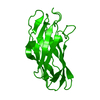
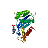
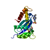
- PDB-3fnk: Crystal structure of the second type II cohesin module from the c... -
+ Open data

- Basic information
| Entry | Database: PDB / ID: 3fnk | ||||||
|---|---|---|---|---|---|---|---|
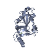
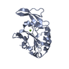
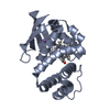
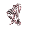
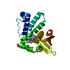
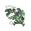
| Title | Crystal structure of the second type II cohesin module from the cellulosomal adaptor ScaA scaffoldin of Acetivibrio cellulolyticus | ||||||
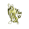
 Components Components | Cellulosomal scaffoldin adaptor protein B | ||||||
 Keywords Keywords | STRUCTURAL PROTEIN / CohB / Dockerin-binding module / beta barrel / alpha helix / beta flaps | ||||||
| Function / homology |  Function and homology information Function and homology informationpolysaccharide catabolic process / hydrolase activity, hydrolyzing O-glycosyl compounds / carbohydrate binding / extracellular region / metal ion binding Similarity search - Function | ||||||
| Biological species |  Acetivibrio cellulolyticus (bacteria) Acetivibrio cellulolyticus (bacteria) | ||||||
| Method |  X-RAY DIFFRACTION / SYNCHROTRON / MOLECULAR REPLACEMENT / Resolution: 1.99 Å X-RAY DIFFRACTION / SYNCHROTRON / MOLECULAR REPLACEMENT / Resolution: 1.99 Å | ||||||
 Authors Authors | Noach, I. / Frolow, F. / Bayer, E.A. | ||||||
 Citation Citation | Journal: J.Mol.Biol. / Year: 2009 Title: Intermodular Linker Flexibility Revealed from Crystal Structures of Adjacent Cellulosomal Cohesins of Acetivibrio cellulolyticus Authors: Noach, I. / Frolow, F. / Alber, O. / Lamed, R. / Shimon, L.J.W. / Bayer, E.A. #1: Journal: Acta Crystallogr.,Sect.F / Year: 2008 Title: Crystallization and preliminary X-ray analysis of Acetivibrio cellulolyticus cellulosomal type II cohesin module: two versions having different linker lengths Authors: Noach, I. / Alber, O. / Bayer, E.A. / Lamed, R. / Levy-Assaraf, M. / Shimon, L.J.W. / Frolow, F. | ||||||
| History |
|
- Structure visualization
| Structure viewer | Molecule: MolmilJmol/JSmol |
|---|
- Downloads & links
-Download
| PDBx/mmCIF format | 3fnk.cif.gz | 127.7 KB | Display | PDBx/mmCIF format |
|---|---|---|---|---|
| PDB format | pdb3fnk.ent.gz | 100.3 KB | Display | PDB format |
| PDBx/mmJSON format | 3fnk.json.gz | Tree view | PDBx/mmJSON format | |
| Others |  Other downloads Other downloads |
-Validation report
| Arichive directory | https://data.pdbj.org/pub/pdb/validation_reports/fn/3fnkftp://data.pdbj.org/pub/pdb/validation_reports/fn/3fnk | HTTPS FTP |
|---|
-Related structure data
| Related structure data | 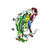 1zv9C  3bwzC  3ghpC  1qznS S: Starting model for refinement C: citing same article ( |
|---|---|
| Similar structure data |


-Links
 PDBj
PDBj


- Assembly
| Deposited unit | 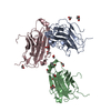
| ||||||||
|---|---|---|---|---|---|---|---|---|---|
| 1 | 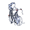
| ||||||||
| 2 | 
| ||||||||
| 3 | 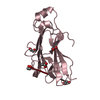
| ||||||||
| Unit cell |
|
-Components
-Protein , 1 types, 3 molecules ABC
| #1: Protein | Mass: 20494.070 Da / Num. of mol.: 3 / Fragment: Cohesin module, UNP residues 190-368 Source method: isolated from a genetically manipulated source Source: (gene. exp.) Acetivibrio cellulolyticus (bacteria) / Gene: scaB / Plasmid: pET28a / Production host: |
|---|
-Non-polymers , 5 types, 557 molecules 

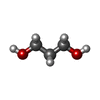
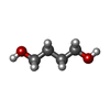

| #2: Chemical | ChemComp-EDO /  Mass: 62.068 Da / Num. of mol.: 8 / Source method: obtained synthetically / Formula: C2H6O2 Mass: 62.068 Da / Num. of mol.: 8 / Source method: obtained synthetically / Formula: C2H6O2#3: Chemical | ChemComp-ACT /  Mass: 59.044 Da / Num. of mol.: 4 / Source method: obtained synthetically / Formula: C2H3O2 Mass: 59.044 Da / Num. of mol.: 4 / Source method: obtained synthetically / Formula: C2H3O2#4: Chemical |  Mass: 76.094 Da / Num. of mol.: 3 / Source method: obtained synthetically / Formula: C3H8O2 Mass: 76.094 Da / Num. of mol.: 3 / Source method: obtained synthetically / Formula: C3H8O2#5: Chemical | ChemComp-BU1 / |  Mass: 90.121 Da / Num. of mol.: 1 / Source method: obtained synthetically / Formula: C4H10O2 Mass: 90.121 Da / Num. of mol.: 1 / Source method: obtained synthetically / Formula: C4H10O2#6: Water | ChemComp-HOH / | Mass: 18.015 Da / Num. of mol.: 541 / Source method: isolated from a natural source / Formula: H2O |
|---|
-Experimental details
-Experiment
| Experiment | Method: X-RAY DIFFRACTION / Number of used crystals: 1 |
|---|
- Sample preparation
| Crystal | Density Matthews: 2.81 Å3/Da / Density % sol: 56.18 % |
|---|---|
| Crystal grow | Temperature: 293 K / Method: vapor diffusion, hanging drop / pH: 4.6 Details: 0.2M ammonium sulfate, 0.1M Sodium acetate trihydrate, 25% PEG 4000, pH 4.6, VAPOR DIFFUSION, HANGING DROP, temperature 293K |
-Data collection
| Diffraction | Mean temperature: 100 K |
|---|---|
| Diffraction source | Source: SYNCHROTRON / Site: ESRF  / Beamline: ID14-3 / Wavelength: 0.9393 Å / Beamline: ID14-3 / Wavelength: 0.9393 Å |
| Detector | Type: ADSC QUANTUM 315 / Detector: CCD / Date: Jun 19, 2006 |
| Radiation | Monochromator: SAGITALLY FOCUSED Si(111) / Protocol: SINGLE WAVELENGTH / Monochromatic (M) / Laue (L): M / Scattering type: x-ray |
| Radiation wavelength | Wavelength: 0.9393 Å / Relative weight: 1 |
| Reflection | Resolution: 1.99→30 Å / Num. all: 47999 / Num. obs: 47999 / % possible obs: 95.5 % / Observed criterion σ(F): 0 / Observed criterion σ(I): 0 / Redundancy: 7.45 % / Biso Wilson estimate: 24 Å2 / Rmerge(I) obs: 0.096 / Rsym value: 0.096 / Net I/σ(I): 19.6 |
| Reflection shell | Resolution: 1.99→2.07 Å / Rmerge(I) obs: 0.48 / Mean I/σ(I) obs: 2.1 / Num. unique all: 3559 / Rsym value: 0.48 / % possible all: 74.9 |
- Processing
| Software |
| |||||||||||||||||||||||||||||||||||||||||||||||||||||||||||||||||
|---|---|---|---|---|---|---|---|---|---|---|---|---|---|---|---|---|---|---|---|---|---|---|---|---|---|---|---|---|---|---|---|---|---|---|---|---|---|---|---|---|---|---|---|---|---|---|---|---|---|---|---|---|---|---|---|---|---|---|---|---|---|---|---|---|---|---|
| Refinement | Method to determine structure: MOLECULAR REPLACEMENT Starting model: PDB entry 1QZN Resolution: 1.99→29.07 Å / Cor.coef. Fo:Fc: 0.956 / Cor.coef. Fo:Fc free: 0.932 / SU B: 3.784 / SU ML: 0.105 / Cross valid method: THROUGHOUT / σ(F): 0 / ESU R: 0.163 / ESU R Free: 0.155 / Stereochemistry target values: MAXIMUM LIKELIHOOD / Details: HYDROGENS HAVE BEEN ADDED IN THE RIDING POSITIONS
| |||||||||||||||||||||||||||||||||||||||||||||||||||||||||||||||||
| Solvent computation | Ion probe radii: 0.8 Å / Shrinkage radii: 0.8 Å / VDW probe radii: 1.4 Å / Solvent model: MASK | |||||||||||||||||||||||||||||||||||||||||||||||||||||||||||||||||
| Displacement parameters | Biso mean: 31.01 Å2
| |||||||||||||||||||||||||||||||||||||||||||||||||||||||||||||||||
| Refine analyze | Luzzati coordinate error obs: 0.215 Å | |||||||||||||||||||||||||||||||||||||||||||||||||||||||||||||||||
| Refinement step | Cycle: LAST / Resolution: 1.99→29.07 Å
| |||||||||||||||||||||||||||||||||||||||||||||||||||||||||||||||||
| Refine LS restraints |
| |||||||||||||||||||||||||||||||||||||||||||||||||||||||||||||||||
| LS refinement shell | Resolution: 1.991→2.043 Å / Total num. of bins used: 20
|
