 Movie
Movie Controller

[English] 日本語
 Yorodumi
- PDB-3zmj: Structure of E.coli rhomboid protease GlpG in complex with monoba... -
+ Open data

- Basic information
| Entry | Database: PDB / ID: 3zmj | ||||||
|---|---|---|---|---|---|---|---|
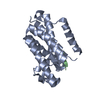
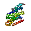
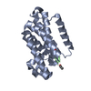
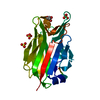
| Title | Structure of E.coli rhomboid protease GlpG in complex with monobactam L61 | ||||||
 Components Components | RHOMBOID PROTEASE GLPG | ||||||
 Keywords Keywords | HYDROLASE / INTRA-MEMBRANE PROTEASE / ACYL ENZYME / BETA LACTAMS / ANITBIOTIC | ||||||
| Function / homology |  Function and homology information Function and homology informationrhomboid protease / endopeptidase activity / serine-type endopeptidase activity / proteolysis / identical protein binding / plasma membrane Similarity search - Function | ||||||
| Biological species |  | ||||||
| Method |  X-RAY DIFFRACTION / SYNCHROTRON / MOLECULAR REPLACEMENT / Resolution: 2.3 Å X-RAY DIFFRACTION / SYNCHROTRON / MOLECULAR REPLACEMENT / Resolution: 2.3 Å | ||||||
 Authors Authors | Vinothkumar, K.R. / Pierrat, O. / Large, J.M. / Freeman, M. | ||||||
 Citation Citation | Journal: Structure / Year: 2013 Title: Structure of Rhomboid Protease in Complex with Beta-Lactam Inhibitors Defines the S2' Cavity. Authors: Vinothkumar, K.R. / Pierrat, O.A. / Large, J.M. / Freeman, M. | ||||||
| History |
|
- Structure visualization
| Structure viewer | Molecule: MolmilJmol/JSmol |
|---|
- Downloads & links
-Download
| PDBx/mmCIF format | 3zmj.cif.gz | 52 KB | Display | PDBx/mmCIF format |
|---|---|---|---|---|
| PDB format | pdb3zmj.ent.gz | 36.1 KB | Display | PDB format |
| PDBx/mmJSON format | 3zmj.json.gz | Tree view | PDBx/mmJSON format | |
| Others |  Other downloads Other downloads |
-Validation report
| Arichive directory | https://data.pdbj.org/pub/pdb/validation_reports/zm/3zmjftp://data.pdbj.org/pub/pdb/validation_reports/zm/3zmj | HTTPS FTP |
|---|
-Related structure data
| Related structure data |  3zmhC  3zmiC  3zotC 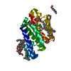 2xovS C: citing same article ( S: Starting model for refinement |
|---|---|
| Similar structure data |

-Links
 PDBj
PDBj
- Assembly
| Deposited unit | 
| ||||||||
|---|---|---|---|---|---|---|---|---|---|
| 1 |
| ||||||||
| Unit cell |
|
-Components
| #1: Protein | Mass: 20214.020 Da / Num. of mol.: 1 / Fragment: CORE TM DOMAIN, RESIDUES 92-270 Source method: isolated from a genetically manipulated source Details: THE BETA LACTAM RING IS OPENED BY THE NUCLEOPHILIC ATTACK OF S201 ON C2-O1 TO FORM A COVALENT BOND Source: (gene. exp.) | ||||||||
|---|---|---|---|---|---|---|---|---|---|
| #2: Chemical | ChemComp-L61 /   Mass: 249.306 Da / Num. of mol.: 1 / Source method: obtained synthetically / Formula: C14H19NO3 Mass: 249.306 Da / Num. of mol.: 1 / Source method: obtained synthetically / Formula: C14H19NO3 | ||||||||
| #3: Sugar | ChemComp-BNG /   Type: D-saccharide / Mass: 306.395 Da / Num. of mol.: 5 Type: D-saccharide / Mass: 306.395 Da / Num. of mol.: 5Source method: isolated from a genetically manipulated source Formula: C15H30O6 / Comment: detergent*YM #4: Chemical |   Mass: 35.453 Da / Num. of mol.: 3 / Source method: obtained synthetically / Formula: Cl Mass: 35.453 Da / Num. of mol.: 3 / Source method: obtained synthetically / Formula: Cl#5: Water | ChemComp-HOH / |  Mass: 18.015 Da / Num. of mol.: 27 / Source method: isolated from a natural source / Formula: H2O Mass: 18.015 Da / Num. of mol.: 27 / Source method: isolated from a natural source / Formula: H2OHas protein modification | Y | Nonpolymer details | ISOBUTYL(1-PHENYLBUT-3-EN-1-YL)CARBAMATE (L61): L61 IS COVALENTLY BONDED TO S201. BNG 404 IS IN ...ISOBUTYL(1-PHENYLBUT-3-EN-1-YL)CARBAMATE (L61): L61 IS COVALENTLY | |
-Experimental details
-Experiment
| Experiment | Method: X-RAY DIFFRACTION / Number of used crystals: 1 |
|---|
- Sample preparation
| Crystal | Density Matthews: 3.5 Å3/Da / Density % sol: 65.6 % / Description: NONE |
|---|---|
| Crystal grow | Temperature: 298 K / Method: vapor diffusion, hanging drop / pH: 7 Details: 1.75 M AMMONIUM CHLORIDE, 0.1M BIS-TRIS, PH 7.0, 298 K |
-Data collection
| Diffraction | Mean temperature: 100 K |
|---|---|
| Diffraction source | Source: SYNCHROTRON / Site: Diamond  / Beamline: I02 / Wavelength: 0.9795 / Beamline: I02 / Wavelength: 0.9795 |
| Detector | Type: ADSC CCD / Detector: CCD / Date: Aug 7, 2011 |
| Radiation | Protocol: SINGLE WAVELENGTH / Monochromatic (M) / Laue (L): M / Scattering type: x-ray |
| Radiation wavelength | Wavelength: 0.9795 Å / Relative weight: 1 |
| Reflection | Resolution: 2.3→53.24 Å / Num. obs: 13419 / % possible obs: 99.2 % / Redundancy: 5.2 % / Biso Wilson estimate: 40.9 Å2 / Rmerge(I) obs: 0.07 / Net I/σ(I): 14.4 |
| Reflection shell | Resolution: 2.3→2.42 Å / Redundancy: 5.2 % / Rmerge(I) obs: 0.59 / Mean I/σ(I) obs: 3 / % possible all: 99.9 |
- Processing
| Software |
| ||||||||||||||||||||||||||||||||||||||||||
|---|---|---|---|---|---|---|---|---|---|---|---|---|---|---|---|---|---|---|---|---|---|---|---|---|---|---|---|---|---|---|---|---|---|---|---|---|---|---|---|---|---|---|---|
| Refinement | Method to determine structure: MOLECULAR REPLACEMENT Starting model: PDB ENTRY 2XOV Resolution: 2.3→53.235 Å / SU ML: 0.28 / σ(F): 1.37 / Phase error: 23.18 / Stereochemistry target values: ML
| ||||||||||||||||||||||||||||||||||||||||||
| Solvent computation | Shrinkage radii: 0.9 Å / VDW probe radii: 1.11 Å / Solvent model: FLAT BULK SOLVENT MODEL | ||||||||||||||||||||||||||||||||||||||||||
| Displacement parameters | Biso mean: 42 Å2 | ||||||||||||||||||||||||||||||||||||||||||
| Refinement step | Cycle: LAST / Resolution: 2.3→53.235 Å
| ||||||||||||||||||||||||||||||||||||||||||
| Refine LS restraints |
| ||||||||||||||||||||||||||||||||||||||||||
| LS refinement shell |
|
