 Movie
Movie Controller


+ Open data

- Basic information
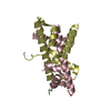
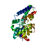
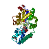
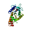
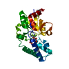
| Entry | Database: PDB / ID: 3hkm | ||||||
|---|---|---|---|---|---|---|---|
| Title | Crystal Structure of rice(Oryza sativa) Rrp46 | ||||||
 Components Components | Os03g0854200 protein | ||||||
 Keywords Keywords | HYDROLASE / RNase PH domain / PHOSPHORYLASE | ||||||
| Function / homology |  Function and homology information Function and homology informationDNA nuclease activity / cytoplasmic exosome (RNase complex) / U4 snRNA 3'-end processing / poly(A)-dependent snoRNA 3'-end processing / nuclear exosome (RNase complex) / nuclear mRNA surveillance / rRNA catabolic process / exonuclease activity / RNA nuclease activity / rRNA processing ...DNA nuclease activity / cytoplasmic exosome (RNase complex) / U4 snRNA 3'-end processing / poly(A)-dependent snoRNA 3'-end processing / nuclear exosome (RNase complex) / nuclear mRNA surveillance / rRNA catabolic process / exonuclease activity / RNA nuclease activity / rRNA processing / double-stranded DNA binding / Hydrolases; Acting on ester bonds; Exoribonucleases producing 5'-phosphomonoesters / single-stranded RNA binding / nucleolus / RNA binding / identical protein binding Similarity search - Function | ||||||
| Biological species |  | ||||||
| Method |  X-RAY DIFFRACTION / SYNCHROTRON / MOLECULAR REPLACEMENT / Resolution: 1.9845 Å X-RAY DIFFRACTION / SYNCHROTRON / MOLECULAR REPLACEMENT / Resolution: 1.9845 Å | ||||||
 Authors Authors | Yang, C.-C. / Wang, Y.-T. / Hsiao, Y.-Y. / Doudeva, L.G. / Yuan, H.S. | ||||||
 Citation Citation | Journal: Rna / Year: 2010 Title: Structural and biochemical characterization of CRN-5 and Rrp46: an exosome component participating in apoptotic DNA degradation Authors: Yang, C.-C. / Wang, Y.-T. / Hsiao, Y.-Y. / Doudeva, L.G. / Kuo, P.-H. / Chow, S.Y. / Yuan, H.S. | ||||||
| History |
|
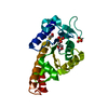
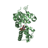
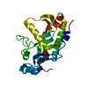
- Structure visualization
| Structure viewer | Molecule: MolmilJmol/JSmol |
|---|
- Downloads & links
-Download
| PDBx/mmCIF format | 3hkm.cif.gz | 135.9 KB | Display | PDBx/mmCIF format |
|---|---|---|---|---|
| PDB format | pdb3hkm.ent.gz | 105.8 KB | Display | PDB format |
| PDBx/mmJSON format | 3hkm.json.gz | Tree view | PDBx/mmJSON format | |
| Others |  Other downloads Other downloads |
-Validation report
| Arichive directory | https://data.pdbj.org/pub/pdb/validation_reports/hk/3hkmftp://data.pdbj.org/pub/pdb/validation_reports/hk/3hkm | HTTPS FTP |
|---|
-Related structure data
| Related structure data |  3krnC 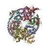 2nn6S S: Starting model for refinement C: citing same article ( |
|---|---|
| Similar structure data |


-Links
 PDBj
PDBj
- Assembly
| Deposited unit | 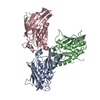
| ||||||||
|---|---|---|---|---|---|---|---|---|---|
| 1 | 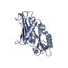
| ||||||||
| 2 | 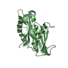
| ||||||||
| 3 | 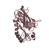
| ||||||||
| Unit cell |
| ||||||||
| Details | DIMER DISSOCIATED INTO MONOMERS IN REMARK 350 THE CRYSTALLIZATION CONDITION |
-Components
| #1: Protein | Mass: 27012.609 Da / Num. of mol.: 3 Source method: isolated from a genetically manipulated source Source: (gene. exp.) Strain: Japonica Group / Gene: putative exonuclease RRP46 / Plasmid: pET22b / Production host:  References: UniProt: Q84T68, Hydrolases; Acting on ester bonds; Exoribonucleases producing 5'-phosphomonoesters #2: Water | ChemComp-HOH / |  Mass: 18.015 Da / Num. of mol.: 416 / Source method: isolated from a natural source / Formula: H2O Mass: 18.015 Da / Num. of mol.: 416 / Source method: isolated from a natural source / Formula: H2O |
|---|
-Experimental details
-Experiment
| Experiment | Method: X-RAY DIFFRACTION / Number of used crystals: 1 |
|---|
- Sample preparation
| Crystal | Density Matthews: 2.56 Å3/Da / Density % sol: 51.96 % Description: The original data was scaled to 2.0 Angstrom by HKL2000. Then refinement was performed with all scaled data automatically by phenix.refine. The highest resolution of 1.98A was made by phenix.refine. |
|---|---|
| Crystal grow | Temperature: 298 K / Method: vapor diffusion, hanging drop / pH: 7 Details: 0.2M sodium malonate, 20% PEG 3350, pH 7.0, VAPOR DIFFUSION, HANGING DROP, temperature 298K |
-Data collection
| Diffraction | Mean temperature: 113 K |
|---|---|
| Diffraction source | Source: SYNCHROTRON / Site: NSRRC  / Beamline: BL13C1 / Wavelength: 1 Å / Beamline: BL13C1 / Wavelength: 1 Å |
| Detector | Type: MAR scanner 345 mm plate / Detector: IMAGE PLATE / Date: Jun 15, 2008 |
| Radiation | Monochromator: Si 111 chnnel / Protocol: SINGLE WAVELENGTH / Monochromatic (M) / Laue (L): M / Scattering type: x-ray |
| Radiation wavelength | Wavelength: 1 Å / Relative weight: 1 |
| Reflection twin | Operator: h,-h-k,-l / Fraction: 0.501 |
| Reflection | Resolution: 1.98→50 Å / Num. obs: 55506 / % possible obs: 100 % / Observed criterion σ(F): 0 / Observed criterion σ(I): 0 / Redundancy: 3.34 % / Rmerge(I) obs: 0.057 / Rsym value: 0.065 / Net I/σ(I): 21.65 / Num. measured all: 185470 |
| Reflection shell | Resolution: 1.98→2.07 Å / Redundancy: 3 % / Rmerge(I) obs: 0.381 / Mean I/σ(I) obs: 3.15 / Num. unique all: 5536 / Rsym value: 0.354 / % possible all: 100 |
- Processing
| Software |
| |||||||||||||||||||||||||||||||||||||||||||||||||||||||||||||||||||||||||||||||||||||||||||||||||||||||||||||||||||||||||||||||||||||||||||||||||||
|---|---|---|---|---|---|---|---|---|---|---|---|---|---|---|---|---|---|---|---|---|---|---|---|---|---|---|---|---|---|---|---|---|---|---|---|---|---|---|---|---|---|---|---|---|---|---|---|---|---|---|---|---|---|---|---|---|---|---|---|---|---|---|---|---|---|---|---|---|---|---|---|---|---|---|---|---|---|---|---|---|---|---|---|---|---|---|---|---|---|---|---|---|---|---|---|---|---|---|---|---|---|---|---|---|---|---|---|---|---|---|---|---|---|---|---|---|---|---|---|---|---|---|---|---|---|---|---|---|---|---|---|---|---|---|---|---|---|---|---|---|---|---|---|---|---|---|---|---|
| Refinement | Method to determine structure: MOLECULAR REPLACEMENT Starting model: PDB entry 2NN6 chain D Resolution: 1.9845→26.807 Å / Occupancy max: 1 / Occupancy min: 1 / FOM work R set: 0.835 / Isotropic thermal model: isotropic / σ(F): 0.12 / Phase error: 24.27 / Stereochemistry target values: TWIN_LSQ_F
| |||||||||||||||||||||||||||||||||||||||||||||||||||||||||||||||||||||||||||||||||||||||||||||||||||||||||||||||||||||||||||||||||||||||||||||||||||
| Solvent computation | Shrinkage radii: 0.9 Å / VDW probe radii: 1.11 Å / Solvent model: FLAT BULK SOLVENT MODEL / Bsol: 51.857 Å2 / ksol: 0.349 e/Å3 | |||||||||||||||||||||||||||||||||||||||||||||||||||||||||||||||||||||||||||||||||||||||||||||||||||||||||||||||||||||||||||||||||||||||||||||||||||
| Displacement parameters | Biso max: 45.5 Å2 / Biso mean: 26.089 Å2 / Biso min: 13.88 Å2
| |||||||||||||||||||||||||||||||||||||||||||||||||||||||||||||||||||||||||||||||||||||||||||||||||||||||||||||||||||||||||||||||||||||||||||||||||||
| Refinement step | Cycle: LAST / Resolution: 1.9845→26.807 Å
| |||||||||||||||||||||||||||||||||||||||||||||||||||||||||||||||||||||||||||||||||||||||||||||||||||||||||||||||||||||||||||||||||||||||||||||||||||
| Refine LS restraints |
| |||||||||||||||||||||||||||||||||||||||||||||||||||||||||||||||||||||||||||||||||||||||||||||||||||||||||||||||||||||||||||||||||||||||||||||||||||
| LS refinement shell |
|
