 Movie
Movie Controller

[English] 日本語
 Yorodumi
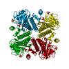
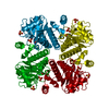
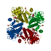
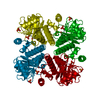
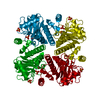
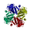
- PDB-1e47: L-Fuculose 1-Phosphate Aldolase from Escherichia coli Mutant E73Q -
+ Open data

- Basic information
| Entry | Database: PDB / ID: 1.0E+47 | |||||||||
|---|---|---|---|---|---|---|---|---|---|---|
| Title | L-Fuculose 1-Phosphate Aldolase from Escherichia coli Mutant E73Q | |||||||||
 Components Components | L-FUCULOSE 1-PHOSPHATE ALDOLASE | |||||||||
 Keywords Keywords | ALDOLASE (CLASS II) / BACTERIAL L-FUCOSE METABOLISM / CLEAVAGE OF L-FUCULOSE 1-PHOSPHATE TO DIHYDROXYACETONE PHOSPHATE AND L-LACTALDEHYDE / MUTANT STRUCTURE | |||||||||
| Function / homology |  Function and homology information Function and homology informationL-fuculose-phosphate aldolase / L-fuculose-phosphate aldolase activity / D-arabinose catabolic process / pentose catabolic process / L-fucose catabolic process / aldehyde-lyase activity / zinc ion binding / cytosol Similarity search - Function | |||||||||
| Biological species |  | |||||||||
| Method |  X-RAY DIFFRACTION / OTHER / Resolution: 2.15 Å X-RAY DIFFRACTION / OTHER / Resolution: 2.15 Å | |||||||||
 Authors Authors | Joerger, A.C. / Schulz, G.E. | |||||||||
 Citation Citation | Journal: J. Mol. Biol. / Year: 2000 Title: Structures of l-fuculose-1-phosphate aldolase mutants outlining motions during catalysis. Authors: Joerger, A.C. / Mueller-Dieckmann, C. / Schulz, G.E. #1: Journal: Biochemistry / Year: 2000Title: Catalytic Action of Fuculose 1-Phosphate Aldolase (Class II) as Derived by Structure-Directed Mutagenesis Authors: Joerger, A.C. / Gosse, C. / Fessner, W.-D. / Schulz, G.E. #2: Journal: J.Mol.Biol. / Year: 1996Title: Catalytic Mechanism of the Metal-Dependent Fuculose Aldolase from Escherichia Coli as Derived from the Structure Authors: Dreyer, M.K. / Schulz, G.E. #3: Journal: J.Mol.Biol. / Year: 1993 Title: The Spatial Structure of the Class II L-Fuculose-1-Phosphate Aldolase from Escherichia Coli Authors: Dreyer, M.K. / Schulz, G.E. | |||||||||
| History |
|
- Structure visualization
| Structure viewer | Molecule: MolmilJmol/JSmol |
|---|
- Downloads & links
-Download
| PDBx/mmCIF format | 1e47.cif.gz | 57.9 KB | Display | PDBx/mmCIF format |
|---|---|---|---|---|
| PDB format | pdb1e47.ent.gz | 41.4 KB | Display | PDB format |
| PDBx/mmJSON format | 1e47.json.gz | Tree view | PDBx/mmJSON format | |
| Others |  Other downloads Other downloads |
-Validation report
| Arichive directory | https://data.pdbj.org/pub/pdb/validation_reports/e4/1e47ftp://data.pdbj.org/pub/pdb/validation_reports/e4/1e47 | HTTPS FTP |
|---|
-Related structure data
| Related structure data |  1e46C  1e48C  1e49C  1e4aC  1e4bC  1e4cC C: citing same article ( |
|---|---|
| Similar structure data |


-Links
 PDBj
PDBj


- Assembly
| Deposited unit | 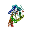
| ||||||||
|---|---|---|---|---|---|---|---|---|---|
| 1 | 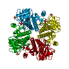
| ||||||||
| Unit cell |
| ||||||||
| Details | BIOMOLECULE |
-Components
-Protein , 1 types, 1 molecules P
| #1: Protein | Mass: 23804.334 Da / Num. of mol.: 1 / Mutation: YES Source method: isolated from a genetically manipulated source Source: (gene. exp.) Description: E73Q MUTATION PERFORMED WITH PHOSPHOROTHIOATE METHOD USING M13MP19 Plasmid: PKKFA2-E73Q / Production host: References: UniProt: P11550, UniProt: P0AB87*PLUS, L-fuculose-phosphate aldolase |
|---|
-Non-polymers , 5 types, 122 molecules 


| #2: Chemical |  Mass: 96.063 Da / Num. of mol.: 2 / Source method: obtained synthetically / Formula: SO4 Mass: 96.063 Da / Num. of mol.: 2 / Source method: obtained synthetically / Formula: SO4#3: Chemical | ChemComp-BME / |  Mass: 78.133 Da / Num. of mol.: 1 / Source method: obtained synthetically / Formula: C2H6OS Mass: 78.133 Da / Num. of mol.: 1 / Source method: obtained synthetically / Formula: C2H6OS#4: Chemical | ChemComp-13P / |  Mass: 170.058 Da / Num. of mol.: 1 / Source method: obtained synthetically / Formula: C3H7O6P Mass: 170.058 Da / Num. of mol.: 1 / Source method: obtained synthetically / Formula: C3H7O6P#5: Chemical | ChemComp-ZN / |  Mass: 65.409 Da / Num. of mol.: 1 / Source method: obtained synthetically / Formula: Zn Mass: 65.409 Da / Num. of mol.: 1 / Source method: obtained synthetically / Formula: Zn#6: Water | ChemComp-HOH / | Mass: 18.015 Da / Num. of mol.: 117 / Source method: isolated from a natural source / Formula: H2O |
|---|
-Details
| Compound details | CHAIN P ENGINEERED| Has protein modification | N | |
|---|
-Experimental details
-Experiment
| Experiment | Method: X-RAY DIFFRACTION / Number of used crystals: 1 |
|---|
- Sample preparation
| Crystal | Density Matthews: 2 Å3/Da / Density % sol: 39 % | ||||||||||||||||||||||||||||||||||||||||||||||||||||||
|---|---|---|---|---|---|---|---|---|---|---|---|---|---|---|---|---|---|---|---|---|---|---|---|---|---|---|---|---|---|---|---|---|---|---|---|---|---|---|---|---|---|---|---|---|---|---|---|---|---|---|---|---|---|---|---|
| Crystal grow | Method: vapor diffusion, hanging drop / pH: 8 Details: CRYSTALS GROWN FROM AMMONIUM SULFATE AT PH 8.0, VAPOUR DIFFUSION, HANGING DROP, CONDITIONS CLOSE TO THE ONES REPORTED FOR THE WILD-TYPE, SEE PDB ID 1FUA FOR FURTHER DETAILS | ||||||||||||||||||||||||||||||||||||||||||||||||||||||
| Crystal grow | *PLUS Temperature: 293 K / pH: 7.6 / Method: vapor diffusion, hanging dropDetails: Dreyer, M.K., (1996) Acta Crystallog. sect., D52, 1082. | ||||||||||||||||||||||||||||||||||||||||||||||||||||||
| Components of the solutions | *PLUS
|
-Data collection
| Diffraction | Mean temperature: 293 K |
|---|---|
| Diffraction source | Source: ROTATING ANODE / Type: RIGAKU RUH2R / Wavelength: 1.5418 |
| Detector | Type: MULTIWIRE SIEMENS X-100 / Detector: AREA DETECTOR / Date: Jul 15, 1997 |
| Radiation | Protocol: SINGLE WAVELENGTH / Monochromatic (M) / Laue (L): M / Scattering type: x-ray |
| Radiation wavelength | Wavelength: 1.5418 Å / Relative weight: 1 |
| Reflection | Resolution: 2.15→10 Å / Num. obs: 10320 / % possible obs: 95 % / Redundancy: 3.4 % / Rsym value: 0.044 / Net I/σ(I): 16 |
| Reflection shell | Resolution: 2.15→2.22 Å / Redundancy: 2.1 % / Mean I/σ(I) obs: 6 / Rsym value: 0.13 / % possible all: 87 |
| Reflection | *PLUS Rmerge(I) obs: 0.044 |
| Reflection shell | *PLUS % possible obs: 87 % / Num. unique obs: 861 / Rmerge(I) obs: 0.13 |
- Processing
| Software |
| ||||||||||||||||||||||||||||||||||||||||||||||||||||||||||||||||||||||||||||||||||||
|---|---|---|---|---|---|---|---|---|---|---|---|---|---|---|---|---|---|---|---|---|---|---|---|---|---|---|---|---|---|---|---|---|---|---|---|---|---|---|---|---|---|---|---|---|---|---|---|---|---|---|---|---|---|---|---|---|---|---|---|---|---|---|---|---|---|---|---|---|---|---|---|---|---|---|---|---|---|---|---|---|---|---|---|---|---|
| Refinement | Method to determine structure: OTHER / Resolution: 2.15→10 Å / SU B: 2.8 / SU ML: 0.07 / σ(F): 0 / ESU R: 0.27 / ESU R Free: 0.19 Details: THE 9 C-TERMINAL RESIDUES (LYS207 - GLU 215) WERE NOT SEEN IN THE DENSITY MAPS
| ||||||||||||||||||||||||||||||||||||||||||||||||||||||||||||||||||||||||||||||||||||
| Displacement parameters | Biso mean: 27.7 Å2 | ||||||||||||||||||||||||||||||||||||||||||||||||||||||||||||||||||||||||||||||||||||
| Refinement step | Cycle: LAST / Resolution: 2.15→10 Å
| ||||||||||||||||||||||||||||||||||||||||||||||||||||||||||||||||||||||||||||||||||||
| Refine LS restraints |
| ||||||||||||||||||||||||||||||||||||||||||||||||||||||||||||||||||||||||||||||||||||
| Software | *PLUS Name: REFMAC / Classification: refinement | ||||||||||||||||||||||||||||||||||||||||||||||||||||||||||||||||||||||||||||||||||||
| Refinement | *PLUS Rfactor obs: 0.15 / Rfactor Rwork: 0.15 | ||||||||||||||||||||||||||||||||||||||||||||||||||||||||||||||||||||||||||||||||||||
| Solvent computation | *PLUS | ||||||||||||||||||||||||||||||||||||||||||||||||||||||||||||||||||||||||||||||||||||
| Displacement parameters | *PLUS |
