[English] 日本語
Yorodumi
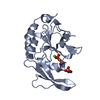
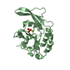
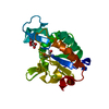
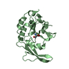
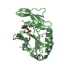
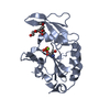
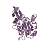
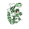
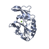
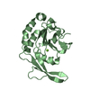
- PDB-3l0y: Crystal structure OF SCP1 phosphatase D98A mutant -

+
Open data


ID or keywords:

Loading...

-
Basic information

Entry
Database: PDB / ID: 3l0y
TitleCrystal structure OF SCP1 phosphatase D98A mutant
ComponentsCarboxy-terminal domain RNA polymerase II polypeptide A small phosphatase 1
KeywordsHYDROLASE / HAD superfamily / Small C-terminal Domain Phosphatase / protein phosphatase
Function / homology
Function and homology information


RNA polymerase II CTD heptapeptide repeat phosphatase activity / myosin phosphatase activity / negative regulation of G1/S transition of mitotic cell cycle / protein-serine/threonine phosphatase / negative regulation of neuron differentiation / protein dephosphorylation / negative regulation of protein phosphorylation / negative regulation of neurogenesis / regulation of transcription by RNA polymerase II / extracellular exosome ...RNA polymerase II CTD heptapeptide repeat phosphatase activity / myosin phosphatase activity / negative regulation of G1/S transition of mitotic cell cycle / protein-serine/threonine phosphatase / negative regulation of neuron differentiation / protein dephosphorylation / negative regulation of protein phosphorylation / negative regulation of neurogenesis / regulation of transcription by RNA polymerase II / extracellular exosome / nucleoplasm / metal ion binding / nucleus
Similarity search - Function
Dullard phosphatase domain, eukaryotic / CTD small RNA polymerase II polypeptide A phosphatase-like / FCP1 homology domain / NLI interacting factor-like phosphatase / FCP1 homology domain profile. / catalytic domain of ctd-like phosphatases / HAD superfamily/HAD-like / HAD superfamily / HAD-like superfamily / Rossmann fold ...Dullard phosphatase domain, eukaryotic / CTD small RNA polymerase II polypeptide A phosphatase-like / FCP1 homology domain / NLI interacting factor-like phosphatase / FCP1 homology domain profile. / catalytic domain of ctd-like phosphatases / HAD superfamily/HAD-like / HAD superfamily / HAD-like superfamily / Rossmann fold / 3-Layer(aba) Sandwich / Alpha Beta
Similarity search - Domain/homology
Carboxy-terminal domain RNA polymerase II polypeptide A small phosphatase 1
Similarity search - Component
Biological speciesHomo sapiens (human)
MethodX-RAY DIFFRACTION / SYNCHROTRON / MOLECULAR REPLACEMENT / Resolution: 2.3 Å
AuthorsZhang, M. / Zhang, Y.
CitationJournal: Protein Sci. / Year: 2010
Title: Structural and functional analysis of the phosphoryl transfer reaction mediated by the human small C-terminal domain phosphatase, Scp1.
Authors: Zhang, M. / Liu, J. / Kim, Y. / Dixon, J.E. / Pfaff, S.L. / Gill, G.N. / Noel, J.P. / Zhang, Y.
History
DepositionDec 10, 2009Deposition site: RCSB / Processing site: RCSB
Revision 1.0Mar 23, 2010Provider: repository / Type: Initial release
Revision 1.1Jul 13, 2011Group: Version format compliance
Revision 1.2Oct 13, 2021Group: Database references / Derived calculations
Category: database_2 / pdbx_struct_conn_angle ...database_2 / pdbx_struct_conn_angle / struct_conn / struct_ref_seq_dif / struct_site
Item: _database_2.pdbx_DOI / _database_2.pdbx_database_accession ..._database_2.pdbx_DOI / _database_2.pdbx_database_accession / _pdbx_struct_conn_angle.ptnr1_auth_comp_id / _pdbx_struct_conn_angle.ptnr1_auth_seq_id / _pdbx_struct_conn_angle.ptnr1_label_atom_id / _pdbx_struct_conn_angle.ptnr1_label_comp_id / _pdbx_struct_conn_angle.ptnr1_label_seq_id / _pdbx_struct_conn_angle.ptnr3_auth_comp_id / _pdbx_struct_conn_angle.ptnr3_auth_seq_id / _pdbx_struct_conn_angle.ptnr3_label_atom_id / _pdbx_struct_conn_angle.ptnr3_label_comp_id / _pdbx_struct_conn_angle.ptnr3_label_seq_id / _pdbx_struct_conn_angle.value / _struct_conn.pdbx_dist_value / _struct_conn.ptnr1_auth_asym_id / _struct_conn.ptnr1_auth_comp_id / _struct_conn.ptnr1_auth_seq_id / _struct_conn.ptnr1_label_asym_id / _struct_conn.ptnr1_label_atom_id / _struct_conn.ptnr1_label_comp_id / _struct_conn.ptnr1_label_seq_id / _struct_conn.ptnr2_auth_asym_id / _struct_conn.ptnr2_auth_comp_id / _struct_conn.ptnr2_auth_seq_id / _struct_conn.ptnr2_label_asym_id / _struct_conn.ptnr2_label_atom_id / _struct_conn.ptnr2_label_comp_id / _struct_ref_seq_dif.details / _struct_site.pdbx_auth_asym_id / _struct_site.pdbx_auth_comp_id / _struct_site.pdbx_auth_seq_id
Revision 1.3Sep 6, 2023Group: Data collection / Refinement description
Category: chem_comp_atom / chem_comp_bond / pdbx_initial_refinement_model

-
Structure visualization

Structure viewerMolecule:
MolmilJmol/JSmol

Downloads & links

-
Assembly

Deposited unit
A: Carboxy-terminal domain RNA polymerase II polypeptide A small phosphatase 1
B: Carboxy-terminal domain RNA polymerase II polypeptide A small phosphatase 1
hetero molecules


Theoretical massNumber of molelcules
Total (without water)42,1184
Polymers42,0702
Non-polymers492
Water2,882160
1
A: Carboxy-terminal domain RNA polymerase II polypeptide A small phosphatase 1
hetero molecules


Theoretical massNumber of molelcules
Total (without water)21,0592
Polymers21,0351
Non-polymers241
Water181
TypeNameSymmetry operationNumber
identity operation1_555x,y,z1
MethodPISA
2
B: Carboxy-terminal domain RNA polymerase II polypeptide A small phosphatase 1
hetero molecules


Theoretical massNumber of molelcules
Total (without water)21,0592
Polymers21,0351
Non-polymers241
Water181
TypeNameSymmetry operationNumber
identity operation1_555x,y,z1
MethodPISA
Unit cell
Length a, b, c (Å)124.791, 78.979, 62.939
Angle α, β, γ (deg.)90.00, 112.79, 90.00
Int Tables number5
Space group name H-MC121
Components on special symmetry positions
IDModelComponents
11A-342-

HOH

-
Components

#1: Protein Carboxy-terminal domain RNA polymerase II polypeptide A small phosphatase 1 / Nuclear LIM interactor-interacting factor 3 / NLI-interacting factor 3 / NLI-IF


Mass: 21034.912 Da / Num. of mol.: 2 / Fragment: UNP residues 77-256 / Mutation: D98A
Source method: isolated from a genetically manipulated source
Source: (gene. exp.) Homo sapiens (human) / Gene: CTDSP1, NIF3, NLIIF, SCP1 / Plasmid: PET28 / Production host: Escherichia coli (E. coli) / Strain (production host): BL21(DE3)
References: UniProt: Q9GZU7, protein-serine/threonine phosphatase
#2: Chemical ChemComp-MG / MAGNESIUM ION


Mass: 24.305 Da / Num. of mol.: 2 / Source method: obtained synthetically / Formula: Mg
#3: Water ChemComp-HOH / water / Water


Mass: 18.015 Da / Num. of mol.: 160 / Source method: isolated from a natural source / Formula: H2O

-
Experimental details

-
Experiment

ExperimentMethod: X-RAY DIFFRACTION / Number of used crystals: 1

-
Sample preparation

CrystalDensity Matthews: 3.4 Å3/Da / Density % sol: 63.81 %
Crystal growTemperature: 298 K / Method: vapor diffusion, sitting drop / pH: 5.5
Details: 0.5M ammonium sulfate, 0.2M lithium sulfate, 100MM HEPES 7.0. Crystal is then transferred to 100MM sodium citrate PH 5.5, 30% PEG8000, 20MM PNPP, 10MM magnesium chloride, VAPOR DIFFUSION, ...Details: 0.5M ammonium sulfate, 0.2M lithium sulfate, 100MM HEPES 7.0. Crystal is then transferred to 100MM sodium citrate PH 5.5, 30% PEG8000, 20MM PNPP, 10MM magnesium chloride, VAPOR DIFFUSION, SITTING DROP, temperature 298.0K

-
Data collection

DiffractionMean temperature: 100 K
Diffraction sourceSource: SYNCHROTRON / Site: ALS / Beamline: 8.2.2 / Wavelength: 1
DetectorType: ADSC QUANTUM 315 / Detector: CCD / Date: May 24, 2006 / Details: MIRRORS
RadiationMonochromator: GRAPHITE / Protocol: SINGLE WAVELENGTH / Monochromatic (M) / Laue (L): M / Scattering type: x-ray
Radiation wavelengthWavelength: 1 Å / Relative weight: 1
ReflectionResolution: 2.3→65.09 Å / Num. obs: 22230 / % possible obs: 90.2 % / Observed criterion σ(I): 0 / Redundancy: 3.4 % / Rsym value: 0.072 / Net I/σ(I): 17.5
Reflection shellResolution: 2.3→2.38 Å / Redundancy: 2.6 % / Mean I/σ(I) obs: 3.4 / Rsym value: 0.205 / % possible all: 57.2

-
Processing

Software
NameVersionClassification
AMoREphasing
REFMAC5.2.0019refinement
HKL-2000data reduction
HKL-2000data scaling
RefinementMethod to determine structure: MOLECULAR REPLACEMENT
Starting model: 2GHQ
Resolution: 2.3→48.98 Å / Cor.coef. Fo:Fc: 0.94 / Cor.coef. Fo:Fc free: 0.919 / SU B: 5.817 / SU ML: 0.142 / Cross valid method: THROUGHOUT / σ(F): 0 / ESU R: 0.276 / ESU R Free: 0.215 / Stereochemistry target values: MAXIMUM LIKELIHOOD / Details: HYDROGENS HAVE BEEN ADDED IN THE RIDING POSITIONS
RfactorNum. reflection% reflectionSelection details
Rfree0.231 1131 5.1 %RANDOM
Rwork0.192 ---
obs0.194 22230 89.8 %-
Solvent computationIon probe radii: 0.8 Å / Shrinkage radii: 0.8 Å / VDW probe radii: 1.4 Å / Solvent model: MASK
Displacement parametersBiso mean: 28.75 Å2
Baniso -1Baniso -2Baniso -3
1-0.4 Å20 Å21.28 Å2
2--1.23 Å20 Å2
3----0.63 Å2
Refinement stepCycle: LAST / Resolution: 2.3→48.98 Å
ProteinNucleic acidLigandSolventTotal
Num. atoms2902 0 2 160 3064
Refine LS restraints
Refine-IDTypeDev idealDev ideal targetNumber
X-RAY DIFFRACTIONr_bond_refined_d0.0110.0222976
X-RAY DIFFRACTIONr_bond_other_d
X-RAY DIFFRACTIONr_angle_refined_deg1.351.9584048
X-RAY DIFFRACTIONr_angle_other_deg
X-RAY DIFFRACTIONr_dihedral_angle_1_deg6.025356
X-RAY DIFFRACTIONr_dihedral_angle_2_deg33.87623.421152
X-RAY DIFFRACTIONr_dihedral_angle_3_deg17.26915486
X-RAY DIFFRACTIONr_dihedral_angle_4_deg22.2511524
X-RAY DIFFRACTIONr_chiral_restr0.0870.2452
X-RAY DIFFRACTIONr_gen_planes_refined0.0050.022304
X-RAY DIFFRACTIONr_gen_planes_other
X-RAY DIFFRACTIONr_nbd_refined0.2090.21190
X-RAY DIFFRACTIONr_nbd_other
X-RAY DIFFRACTIONr_nbtor_refined0.3070.21962
X-RAY DIFFRACTIONr_nbtor_other
X-RAY DIFFRACTIONr_xyhbond_nbd_refined0.1640.2204
X-RAY DIFFRACTIONr_xyhbond_nbd_other
X-RAY DIFFRACTIONr_metal_ion_refined0.0450.21
X-RAY DIFFRACTIONr_metal_ion_other
X-RAY DIFFRACTIONr_symmetry_vdw_refined0.150.239
X-RAY DIFFRACTIONr_symmetry_vdw_other
X-RAY DIFFRACTIONr_symmetry_hbond_refined0.140.213
X-RAY DIFFRACTIONr_symmetry_hbond_other
X-RAY DIFFRACTIONr_symmetry_metal_ion_refined
X-RAY DIFFRACTIONr_symmetry_metal_ion_other
X-RAY DIFFRACTIONr_mcbond_it0.6721.51829
X-RAY DIFFRACTIONr_mcbond_other
X-RAY DIFFRACTIONr_mcangle_it1.20322920
X-RAY DIFFRACTIONr_scbond_it1.76331267
X-RAY DIFFRACTIONr_scangle_it2.8374.51128
X-RAY DIFFRACTIONr_rigid_bond_restr
X-RAY DIFFRACTIONr_sphericity_free
X-RAY DIFFRACTIONr_sphericity_bonded
LS refinement shellResolution: 2.3→2.37 Å / Total num. of bins used: 20
RfactorNum. reflection% reflection
Rfree0.306 54 -
Rwork0.24 880 -
obs--51.69 %

+
About Yorodumi

-
News

-
Feb 9, 2022. New format data for meta-information of EMDB entries

New format data for meta-information of EMDB entries

  • Version 3 of the EMDB header file is now the official format.
  • The previous official version 1.9 will be removed from the archive.

Related info.:EMDB header

External links:wwPDB to switch to version 3 of the EMDB data model

-
Aug 12, 2020. Covid-19 info

Covid-19 info

URL: https://pdbj.org/emnavi/covid19.php

New page: Covid-19 featured information page in EM Navigator.

Related info.:Covid-19 info / Mar 5, 2020. Novel coronavirus structure data

+
Mar 5, 2020. Novel coronavirus structure data

Novel coronavirus structure data

Related info.:Yorodumi Speices / Aug 12, 2020. Covid-19 info

External links:COVID-19 featured content - PDBj / Molecule of the Month (242):Coronavirus Proteases

+
Jan 31, 2019. EMDB accession codes are about to change! (news from PDBe EMDB page)

EMDB accession codes are about to change! (news from PDBe EMDB page)

  • The allocation of 4 digits for EMDB accession codes will soon come to an end. Whilst these codes will remain in use, new EMDB accession codes will include an additional digit and will expand incrementally as the available range of codes is exhausted. The current 4-digit format prefixed with “EMD-” (i.e. EMD-XXXX) will advance to a 5-digit format (i.e. EMD-XXXXX), and so on. It is currently estimated that the 4-digit codes will be depleted around Spring 2019, at which point the 5-digit format will come into force.
  • The EM Navigator/Yorodumi systems omit the EMD- prefix.

Related info.:Q: What is EMD? / ID/Accession-code notation in Yorodumi/EM Navigator

External links:EMDB Accession Codes are Changing Soon! / Contact to PDBj

+
Jul 12, 2017. Major update of PDB

Major update of PDB

  • wwPDB released updated PDB data conforming to the new PDBx/mmCIF dictionary.
  • This is a major update changing the version number from 4 to 5, and with Remediation, in which all the entries are updated.
  • In this update, many items about electron microscopy experimental information are reorganized (e.g. em_software).
  • Now, EM Navigator and Yorodumi are based on the updated data.

External links:wwPDB Remediation / Enriched Model Files Conforming to OneDep Data Standards Now Available in the PDB FTP Archive

-
Yorodumi

Thousand views of thousand structures

  • Yorodumi is a browser for structure data from EMDB, PDB, SASBDB, etc.
  • This page is also the successor to EM Navigator detail page, and also detail information page/front-end page for Omokage search.
  • The word "yorodu" (or yorozu) is an old Japanese word meaning "ten thousand". "mi" (miru) is to see.

Related info.:EMDB / PDB / SASBDB / Comparison of 3 databanks / Yorodumi Search / Aug 31, 2016. New EM Navigator & Yorodumi / Yorodumi Papers / Jmol/JSmol / Function and homology information / Changes in new EM Navigator and Yorodumi

Read more