 Movie
Movie Controller

[English] 日本語
 Yorodumi
- PDB-3epf: CryoEM structure of poliovirus receptor bound to poliovirus type 2 -
+ Open data

- Basic information
| Entry | Database: PDB / ID: 3epf | ||||||
|---|---|---|---|---|---|---|---|
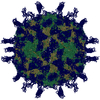
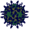
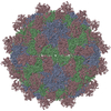
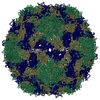
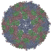
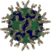
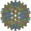
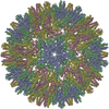
| Title | CryoEM structure of poliovirus receptor bound to poliovirus type 2 | ||||||
 Components Components |
| ||||||
 Keywords Keywords | VIRUS / CD155 structure Immunoglobulin Superfamily / poliovirus capsid jelly role / Cell adhesion / Cell membrane / Glycoprotein / Host-virus interaction / Immunoglobulin domain / Membrane / Receptor / Secreted / Transmembrane / VIRAL PROTEIN | ||||||
| Function / homology |  Function and homology information Function and homology informationsusceptibility to T cell mediated cytotoxicity / susceptibility to natural killer cell mediated cytotoxicity / Nectin/Necl trans heterodimerization / positive regulation of natural killer cell mediated cytotoxicity directed against tumor cell target / cell adhesion mediator activity / positive regulation of natural killer cell mediated cytotoxicity / negative regulation of natural killer cell mediated cytotoxicity / heterophilic cell-cell adhesion / natural killer cell mediated cytotoxicity / homophilic cell-cell adhesion ...susceptibility to T cell mediated cytotoxicity / susceptibility to natural killer cell mediated cytotoxicity / Nectin/Necl trans heterodimerization / positive regulation of natural killer cell mediated cytotoxicity directed against tumor cell target / cell adhesion mediator activity / positive regulation of natural killer cell mediated cytotoxicity / negative regulation of natural killer cell mediated cytotoxicity / heterophilic cell-cell adhesion / natural killer cell mediated cytotoxicity / homophilic cell-cell adhesion / symbiont-mediated suppression of host cytoplasmic pattern recognition receptor signaling pathway via inhibition of RIG-I activity / symbiont-mediated suppression of host cytoplasmic pattern recognition receptor signaling pathway via inhibition of MDA-5 activity / receptor-mediated endocytosis of virus by host cell / symbiont-mediated suppression of host cytoplasmic pattern recognition receptor signaling pathway via inhibition of MAVS activity / cell adhesion molecule binding / picornain 2A / symbiont-mediated suppression of host mRNA export from nucleus / adherens junction / symbiont genome entry into host cell via pore formation in plasma membrane / picornain 3C / T=pseudo3 icosahedral viral capsid / host cell cytoplasmic vesicle membrane / Immunoregulatory interactions between a Lymphoid and a non-Lymphoid cell / ribonucleoside triphosphate phosphatase activity / nucleoside-triphosphate phosphatase / virus receptor activity / channel activity / signaling receptor activity / monoatomic ion transmembrane transport / DNA replication / RNA helicase activity / receptor ligand activity / symbiont-mediated activation of host autophagy / RNA-directed RNA polymerase / cysteine-type endopeptidase activity / viral RNA genome replication / focal adhesion / RNA-directed RNA polymerase activity / virion attachment to host cell / host cell nucleus / DNA-templated transcription / structural molecule activity / cell surface / proteolysis / : / RNA binding / zinc ion binding / ATP binding / membrane / plasma membrane / cytoplasm Similarity search - Function | ||||||
| Biological species |  Homo sapiens (human) Homo sapiens (human) Poliovirus type 2 Poliovirus type 2 | ||||||
| Method | ELECTRON MICROSCOPY / single particle reconstruction / cryo EM / Resolution: 9 Å | ||||||
 Authors Authors | Zhang, P. / Mueller, S. / Morais, M.C. / Bator, C.M. / Bowman, V.D. / Hafenstein, S. / Wimmer, E. / Rossmann, M.G. | ||||||
 Citation Citation | Journal: Proc Natl Acad Sci U S A / Year: 2008 Title: Crystal structure of CD155 and electron microscopic studies of its complexes with polioviruses. Authors: Ping Zhang / Steffen Mueller / Marc C Morais / Carol M Bator / Valorie D Bowman / Susan Hafenstein / Eckard Wimmer / Michael G Rossmann /  Abstract: When poliovirus (PV) recognizes its receptor, CD155, the virus changes from a 160S to a 135S particle before releasing its genome into the cytoplasm. CD155 is a transmembrane protein with 3 Ig-like ...When poliovirus (PV) recognizes its receptor, CD155, the virus changes from a 160S to a 135S particle before releasing its genome into the cytoplasm. CD155 is a transmembrane protein with 3 Ig-like extracellular domains, D1-D3, where D1 is recognized by the virus. The crystal structure of D1D2 has been determined to 3.5-A resolution and fitted into approximately 8.5-A resolution cryoelectron microscopy reconstructions of the virus-receptor complexes for the 3 PV serotypes. These structures show that, compared with human rhinoviruses, the virus-receptor interactions for PVs have a greater dependence on hydrophobic interactions, as might be required for a virus that can inhabit environments of different pH. The pocket factor was shown to remain in the virus during the first recognition stage. The present structures, when combined with earlier mutational investigations, show that in the subsequent entry stage the receptor moves further into the canyon when at a physiological temperature, thereby expelling the pocket factor and separating the viral subunits to form 135S particles. These results provide a detailed analysis of how a nonenveloped virus can enter its host cell. | ||||||
| History |
|
- Structure visualization
| Movie |
Movie viewer |
|---|---|
| Structure viewer | Molecule: MolmilJmol/JSmol |
- Downloads & links
-Download
| PDBx/mmCIF format | 3epf.cif.gz | 226.5 KB | Display | PDBx/mmCIF format |
|---|---|---|---|---|
| PDB format | pdb3epf.ent.gz | 174.2 KB | Display | PDB format |
| PDBx/mmJSON format | 3epf.json.gz | Tree view | PDBx/mmJSON format | |
| Others |  Other downloads Other downloads |
-Validation report
| Arichive directory | https://data.pdbj.org/pub/pdb/validation_reports/ep/3epfftp://data.pdbj.org/pub/pdb/validation_reports/ep/3epf | HTTPS FTP |
|---|
-Related structure data
| Related structure data | 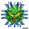 1563MC 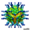 1562C 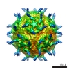 1570C  3epcC  3epdC  3uroC M: map data used to model this data C: citing same article ( |
|---|---|
| Similar structure data |


-Links
 PDBj
PDBj


- Assembly
| Deposited unit | 
|
|---|---|
| 1 | x 60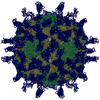
|
| 2 |
|
| 3 | x 5
|
| 4 | x 6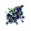
|
| 5 | 
|
| Symmetry | Point symmetry: (Schoenflies symbol: I (icosahedral)) |
-Components
-Protein , 5 types, 5 molecules R1243
| #1: Protein | Mass: 23330.514 Da / Num. of mol.: 1 / Fragment: Poliovirus receptor CD155 D1D2 / Mutation: N105D, N120S, N188Q, N218Q, N237S Source method: isolated from a genetically manipulated source Source: (gene. exp.) Homo sapiens (human) / Gene: PVR, PVS / Production host:  |
|---|---|
| #2: Protein | Mass: 30734.535 Da / Num. of mol.: 1 Source method: isolated from a genetically manipulated source Source: (gene. exp.) Poliovirus type 2 / Production host: |
| #3: Protein | Mass: 29045.814 Da / Num. of mol.: 1 Source method: isolated from a genetically manipulated source Source: (gene. exp.) Poliovirus type 2 / Production host: |
| #4: Protein | Mass: 7346.957 Da / Num. of mol.: 1 Source method: isolated from a genetically manipulated source Source: (gene. exp.) Poliovirus type 2 / Production host: |
| #5: Protein | Mass: 26115.852 Da / Num. of mol.: 1 Source method: isolated from a genetically manipulated source Source: (gene. exp.) Poliovirus type 2 / Production host: |
-Non-polymers , 3 types, 273 molecules 


| #6: Chemical | ChemComp-SC4 /  Mass: 423.717 Da / Num. of mol.: 1 / Source method: obtained synthetically / Formula: C21H17Cl3O3 Mass: 423.717 Da / Num. of mol.: 1 / Source method: obtained synthetically / Formula: C21H17Cl3O3 |
|---|---|
| #7: Chemical | ChemComp-MYR /  Mass: 228.371 Da / Num. of mol.: 1 / Source method: obtained synthetically / Formula: C14H28O2 Mass: 228.371 Da / Num. of mol.: 1 / Source method: obtained synthetically / Formula: C14H28O2 |
| #8: Water | ChemComp-HOH / Mass: 18.015 Da / Num. of mol.: 271 / Source method: isolated from a natural source / Formula: H2O |
-Details
| Has protein modification | Y |
|---|
-Experimental details
-Experiment
| Experiment | Method: ELECTRON MICROSCOPY |
|---|---|
| EM experiment | Aggregation state: PARTICLE / 3D reconstruction method: single particle reconstruction |
- Sample preparation
| Component |
| ||||||||||||||||
|---|---|---|---|---|---|---|---|---|---|---|---|---|---|---|---|---|---|
| Buffer solution | pH: 7.5 / Details: 10mM Tris-HCl, 20mM NaCl | ||||||||||||||||
| Specimen | Embedding applied: NO / Shadowing applied: NO / Staining applied: NO / Vitrification applied: YES | ||||||||||||||||
| Vitrification | Cryogen name: ETHANE |
- Electron microscopy imaging
| Microscopy | Model: FEI/PHILIPS CM300FEG/T |
|---|---|
| Electron gun | Electron source:  FIELD EMISSION GUN / Accelerating voltage: 300 kV / Illumination mode: FLOOD BEAM FIELD EMISSION GUN / Accelerating voltage: 300 kV / Illumination mode: FLOOD BEAM |
| Electron lens | Mode: BRIGHT FIELD / Nominal magnification: 47000 X / Nominal defocus max: 2745 nm / Nominal defocus min: 1236 nm |
| Image recording | Electron dose: 20 e/Å2 / Film or detector model: KODAK SO-163 FILM |
- Processing
| EM software | Name: EMfit / Category: model fitting | ||||||||||||
|---|---|---|---|---|---|---|---|---|---|---|---|---|---|
| Symmetry | Point symmetry: I (icosahedral) | ||||||||||||
| 3D reconstruction | Resolution: 9 Å / Nominal pixel size: 2.69 Å / Actual pixel size: 2.66 Å / Symmetry type: POINT | ||||||||||||
| Atomic model building | Space: REAL | ||||||||||||
| Refinement step | Cycle: LAST
|
