[English] 日本語
Yorodumi
- PDB-3tn9: X-ray structure of the HRV2 empty capsid (B-particle) -

+
Open data


ID or keywords:

Loading...

-
Basic information

Entry
Database: PDB / ID: 3tn9
TitleX-ray structure of the HRV2 empty capsid (B-particle)
Components
  • Protein VP1
  • Protein VP2
  • Protein VP3
KeywordsVIRUS / Empty capsid / B-particle / 80S particle / rhinovirus / HRV2 / uncoating / Rossmann Fold
Function / homology
Function and homology information


symbiont-mediated suppression of host cytoplasmic pattern recognition receptor signaling pathway via inhibition of RIG-I activity / picornain 2A / symbiont-mediated suppression of host mRNA export from nucleus / symbiont genome entry into host cell via pore formation in plasma membrane / picornain 3C / ribonucleoside triphosphate phosphatase activity / T=pseudo3 icosahedral viral capsid / host cell cytoplasmic vesicle membrane / endocytosis involved in viral entry into host cell / : ...symbiont-mediated suppression of host cytoplasmic pattern recognition receptor signaling pathway via inhibition of RIG-I activity / picornain 2A / symbiont-mediated suppression of host mRNA export from nucleus / symbiont genome entry into host cell via pore formation in plasma membrane / picornain 3C / ribonucleoside triphosphate phosphatase activity / T=pseudo3 icosahedral viral capsid / host cell cytoplasmic vesicle membrane / endocytosis involved in viral entry into host cell / : / nucleoside-triphosphate phosphatase / protein complex oligomerization / monoatomic ion channel activity / DNA replication / RNA helicase activity / induction by virus of host autophagy / RNA-directed RNA polymerase / symbiont-mediated suppression of host gene expression / viral RNA genome replication / cysteine-type endopeptidase activity / RNA-dependent RNA polymerase activity / DNA-templated transcription / host cell nucleus / structural molecule activity / virion attachment to host cell / proteolysis / RNA binding / ATP binding / membrane / metal ion binding
Similarity search - Function
Jelly Rolls - #20 / Poliovirus 3A protein-like / Poliovirus 3A protein like / Picornavirus 2B protein / Poliovirus core protein 3a, soluble domain / Picornavirus 2B protein / Peptidase C3, picornavirus core protein 2A / Picornavirus core protein 2A / Picornavirus coat protein VP4 / Picornavirus coat protein (VP4) ...Jelly Rolls - #20 / Poliovirus 3A protein-like / Poliovirus 3A protein like / Picornavirus 2B protein / Poliovirus core protein 3a, soluble domain / Picornavirus 2B protein / Peptidase C3, picornavirus core protein 2A / Picornavirus core protein 2A / Picornavirus coat protein VP4 / Picornavirus coat protein (VP4) / Picornavirales 3C/3C-like protease domain / Picornavirales 3C/3C-like protease domain profile. / Peptidase C3A/C3B, picornaviral / 3C cysteine protease (picornain 3C) / Picornavirus capsid / picornavirus capsid protein / Helicase, superfamily 3, single-stranded RNA virus / Superfamily 3 helicase of positive ssRNA viruses domain profile. / Helicase, superfamily 3, single-stranded DNA/RNA virus / RNA helicase / Picornavirus/Calicivirus coat protein / Viral coat protein subunit / RNA-directed RNA polymerase, C-terminal domain / Viral RNA-dependent RNA polymerase / Reverse transcriptase/Diguanylate cyclase domain / Jelly Rolls / RNA-directed RNA polymerase, catalytic domain / RdRp of positive ssRNA viruses catalytic domain profile. / Peptidase S1, PA clan, chymotrypsin-like fold / DNA/RNA polymerase superfamily / Peptidase S1, PA clan / Sandwich / P-loop containing nucleoside triphosphate hydrolase / Mainly Beta
Similarity search - Domain/homology
Biological speciesHuman rhinovirus 2
MethodX-RAY DIFFRACTION / SYNCHROTRON / RIGID BODY FITTING / Resolution: 3 Å
AuthorsGarriga, D. / Pickl-Herk, A. / Luque, D. / Wruss, J. / Caston, J.R. / Blaas, D. / Verdaguer, N.
CitationJournal: Plos Pathog. / Year: 2012
Title: Insights into minor group rhinovirus uncoating: the X-ray structure of the HRV2 empty capsid.
Authors: Garriga, D. / Pickl-Herk, A. / Luque, D. / Wruss, J. / Caston, J.R. / Blaas, D. / Verdaguer, N.
History
DepositionSep 1, 2011Deposition site: RCSB / Processing site: RCSB
Revision 1.0Mar 28, 2012Provider: repository / Type: Initial release
Revision 1.1Sep 13, 2023Group: Data collection / Database references ...Data collection / Database references / Derived calculations / Refinement description
Category: chem_comp_atom / chem_comp_bond ...chem_comp_atom / chem_comp_bond / database_2 / pdbx_initial_refinement_model / pdbx_struct_oper_list
Item: _database_2.pdbx_DOI / _database_2.pdbx_database_accession ..._database_2.pdbx_DOI / _database_2.pdbx_database_accession / _pdbx_struct_oper_list.name / _pdbx_struct_oper_list.symmetry_operation / _pdbx_struct_oper_list.type

-
Structure visualization

Structure viewerMolecule:
MolmilJmol/JSmol

Downloads & links

-
Assembly

Deposited unit
1: Protein VP1
2: Protein VP2
3: Protein VP3


Theoretical massNumber of molelcules
Total (without water)88,0423
Polymers88,0423
Non-polymers00
Water0
1
1: Protein VP1
2: Protein VP2
3: Protein VP3
x 60


Theoretical massNumber of molelcules
Total (without water)5,282,531180
Polymers5,282,531180
Non-polymers00
Water0
TypeNameSymmetry operationNumber
identity operation1_555x,y,z1
point symmetry operation59
2


  • Idetical with deposited unit
  • icosahedral asymmetric unit
TypeNameSymmetry operationNumber
identity operation1_555x,y,z1
3
1: Protein VP1
2: Protein VP2
3: Protein VP3
x 5


  • icosahedral pentamer
  • 440 kDa, 15 polymers
Theoretical massNumber of molelcules
Total (without water)440,21115
Polymers440,21115
Non-polymers00
Water0
TypeNameSymmetry operationNumber
identity operation1_555x,y,z1
point symmetry operation4
4
1: Protein VP1
2: Protein VP2
3: Protein VP3
x 6


  • icosahedral 23 hexamer
  • 528 kDa, 18 polymers
Theoretical massNumber of molelcules
Total (without water)528,25318
Polymers528,25318
Non-polymers00
Water0
TypeNameSymmetry operationNumber
identity operation1_555x,y,z1
point symmetry operation5
5


  • Idetical with deposited unit in distinct coordinate
  • icosahedral asymmetric unit, std point frame
TypeNameSymmetry operationNumber
transform to point frame1
6
1: Protein VP1
2: Protein VP2
3: Protein VP3
x 15


  • crystal asymmetric unit, crystal frame
  • 1.32 MDa, 45 polymers
Theoretical massNumber of molelcules
Total (without water)1,320,63345
Polymers1,320,63345
Non-polymers00
Water0
TypeNameSymmetry operationNumber
identity operation1_555x,y,z2
point symmetry operation14
Unit cell
Length a, b, c (Å)314.010, 356.850, 382.470
Angle α, β, γ (deg.)90.000, 90.000, 90.000
Int Tables number23
Space group name H-MI222
SymmetryPoint symmetry: (Schoenflies symbol: I (icosahedral))
Noncrystallographic symmetry (NCS)NCS oper:
IDCodeMatrix
1given(1), (1), (1)
2generate(0.30901699, -0.80901699, -0.5), (0.809017, 0.5, -0.30901699), (0.5, -0.309017, 0.80901699)
3generate(-0.809017, -0.5, -0.30901699), (0.5, -0.30901699, -0.80901699), (0.30901699, -0.809017, 0.5)
4generate(-0.80901699, 0.5, 0.309017), (-0.5, -0.309017, -0.80901699), (-0.309017, -0.80901699, 0.5)
5generate(0.309017, 0.80901699, 0.5), (-0.80901699, 0.5, -0.30901699), (-0.5, -0.30901699, 0.809017)
6generate(-0.80901699, -0.50000001, 0.309017), (-0.49999999, 0.30901699, -0.809017), (0.30901699, -0.80901699, -0.5)
7generate(-0.5, 0.30901699, 0.809017), (-0.30901699, 0.80901699, -0.5), (-0.809017, -0.5, -0.30901699)
8generate(0.5, 0.30901699, 0.80901699), (0.30901699, 0.809017, -0.5), (-0.80901699, 0.5, 0.309017)
9generate(0.80901699, -0.5, 0.309017), (0.5, 0.309017, -0.80901699), (0.309017, 0.80901699, 0.5)
10generate(-1), (-1), (1)
11generate(-0.309017, -0.80901699, 0.50000001), (0.809017, -0.49999999, -0.30901699), (0.5, 0.30901699, 0.80901699)
12generate(-0.5, -0.309017, 0.809017), (-0.30901699, -0.809017, -0.5), (0.80901699, -0.5, 0.309017)
13generate(1), (-1), (-1)
14generate(0.5, -0.309017, 0.80901699), (-0.309017, 0.80901699, 0.5), (-0.80901699, -0.5, 0.309017)
15generate(0.30901699, -0.809017, 0.5), (0.80901699, 0.5, 0.30901699), (-0.5, 0.30901699, 0.809017)
Details
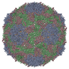
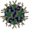
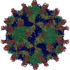
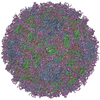
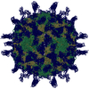
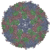
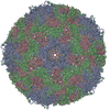
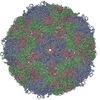
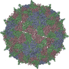
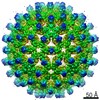
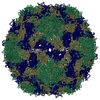
biological unit is the complete icosahedral capsid, generated by 60 protomers (apply matrices from REMARK 350)

-
Components

#1: Protein Protein VP1 / P1D / Virion protein 1


Mass: 32924.797 Da / Num. of mol.: 1 / Source method: isolated from a natural source
Details: Virions were propagated in HeLaH1 cells. Empty particles were obtained by heating to 56 C for 12 minutes.
Source: (natural) Human rhinovirus 2 / Strain: HRV2 / References: UniProt: P04936
#2: Protein Protein VP2 / P1B / Virion protein 2


Mass: 29009.588 Da / Num. of mol.: 1 / Source method: isolated from a natural source
Details: Virions were propagated in HeLaH1 cells. Empty particles were obtained by heating to 56 C for 12 minutes.
Source: (natural) Human rhinovirus 2 / Strain: HRV2 / References: UniProt: P04936
#3: Protein Protein VP3 / P1C / Virion protein 3


Mass: 26107.793 Da / Num. of mol.: 1 / Source method: isolated from a natural source
Details: Virions were propagated in HeLaH1 cells. Empty particles were obtained by heating to 56 C for 12 minutes.
Source: (natural) Human rhinovirus 2 / Strain: HRV2 / References: UniProt: P04936

-
Experimental details

-
Experiment

ExperimentMethod: X-RAY DIFFRACTION / Number of used crystals: 10

-
Sample preparation

Crystal growTemperature: 293 K / Method: vapor diffusion, hanging drop / pH: 6.5
Details: 0.1M sodium acetate pH 6.5 - 8.0, 5% glycerol, VAPOR DIFFUSION, HANGING DROP, temperature 293K

-
Data collection

Diffraction
IDMean temperature (K)Crystal-ID
11001
21001
Diffraction source
SourceSiteBeamlineIDWavelength (Å)
SYNCHROTRONESRF ID23-110.979
SYNCHROTRONSLS X06DA21.006
Detector
TypeIDDetectorDate
ADSC QUANTUM 315r1CCDOct 27, 2008
MARMOSAIC 225 mm CCD2CCDJan 30, 2009
Radiation
IDMonochromatorProtocolMonochromatic (M) / Laue (L)Scattering typeWavelength-ID
1Si 111 CHANNELSINGLE WAVELENGTHMx-ray1
2DCCMSINGLE WAVELENGTHMx-ray1
Radiation wavelength
IDWavelength (Å)Relative weight
10.9791
21.0061
ReflectionResolution: 2.993→260.92 Å / Num. all: 308652 / Num. obs: 308652 / % possible obs: 72.7 % / Observed criterion σ(F): 1 / Observed criterion σ(I): 1 / Redundancy: 3.2 % / Rmerge(I) obs: 0.199 / Rsym value: 0.199 / Net I/σ(I): 5.7
Reflection shell

Diffraction-ID: 1,2

Resolution (Å)Redundancy (%)Rmerge(I) obsMean I/σ(I) obsNum. measured allNum. unique allRsym value% possible all
3-3.151.30.4511.429881231150.45140.3
3.15-3.341.50.391.847871320960.3954.4
3.34-3.571.70.3292.157627341190.32961.5
3.57-3.862.20.2931.987196403130.29377.9
3.86-4.233.20.272.1131699412080.2786.4
4.23-4.724.10.2223.1157674386580.22289.5
4.72-5.464.30.1913.5149214343780.19190
5.46-6.684.60.1843.7132581289500.18489.6
6.68-9.455.10.1514.1118950231090.15192
9.45-60.7465.40.1354.168060127060.13590.5

-
Phasing

Phasing dmMethod: Solvent flattening / Reflection: 257725
Phasing dm shell
Resolution (Å)Delta phi finalFOM Reflection
17.89-10056.70.712535
12.65-17.8942.50.9043443
10.33-12.6539.30.9454460
8.94-10.3338.60.955265
8-8.9444.50.9455973
7.3-844.40.9396539
6.76-7.3440.9386996
6.32-6.7641.80.9347414
5.96-6.32380.9367847
5.66-5.9641.30.9348298
5.39-5.6640.60.9278724
5.16-5.3938.90.9259108
4.96-5.1637.90.929563
4.78-4.9636.90.9189921
4.62-4.7839.80.9110252
4.47-4.6239.60.90210621
4.34-4.47390.90810837
4.22-4.3440.20.90611105
4.1-4.2240.70.90211409
4-4.141.10.89311696
3.9-4420.87611369
3.81-3.939.20.86911253
3.73-3.8139.50.88511210
3.65-3.7338.90.86211358
3.58-3.6538.40.86311152
3.51-3.5838.60.87210850
3.44-3.5140.40.8649382
3.38-3.4442.70.8569048
3.3-3.3847.20.75512097

-
Processing

Software
NameVersionClassificationNB
MOSFLMdata reduction
SCALA3.2.25data scaling
DM6.1phasing
CNSrefinement
PDB_EXTRACT3.1data extraction
DNAdata collection
CNSphasing
RefinementMethod to determine structure: RIGID BODY FITTING
Starting model: Native HRV2 structure PDB entry 1FPN
Resolution: 3→50 Å / Occupancy max: 1 / Occupancy min: 0 / FOM work R set: 0.7771 / Isotropic thermal model: isotropic / σ(F): 0 / Stereochemistry target values: Engh & Huber
Details: 15-fold non-crystallographic symmetry constraints were used during refinement.
RfactorNum. reflection% reflectionSelection details
Rfree0.2646 15275 3.6 %RANDOM, 5%
Rwork0.2654 ---
all0.2654 307962 --
obs0.2654 307962 72.7 %-
Solvent computationBsol: 9.454 Å2
Displacement parametersBiso max: 140.79 Å2 / Biso mean: 50.6204 Å2 / Biso min: 26.58 Å2
Refinement stepCycle: LAST / Resolution: 3→50 Å
ProteinNucleic acidLigandSolventTotal
Num. atoms5516 0 0 0 5516
Refine LS restraints
Refine-IDTypeDev idealDev ideal target
X-RAY DIFFRACTIONc_mcbond_it1.2751.5
X-RAY DIFFRACTIONc_scbond_it1.362
X-RAY DIFFRACTIONc_mcangle_it2.3722
X-RAY DIFFRACTIONc_scangle_it2.3132.5
Xplor file
Refine-IDSerial noParam file
X-RAY DIFFRACTION1CNS_TOPPAR:protein_rep.param
X-RAY DIFFRACTION2CNS_TOPPAR:dna-rna_rep.param
X-RAY DIFFRACTION3CNS_TOPPAR:water_rep.param
X-RAY DIFFRACTION4CNS_TOPPAR:ion.param
X-RAY DIFFRACTION5CNS_TOPPAR:carbohydrate.param

+
About Yorodumi

-
News

-
Feb 9, 2022. New format data for meta-information of EMDB entries

New format data for meta-information of EMDB entries

  • Version 3 of the EMDB header file is now the official format.
  • The previous official version 1.9 will be removed from the archive.

Related info.:EMDB header

External links:wwPDB to switch to version 3 of the EMDB data model

-
Aug 12, 2020. Covid-19 info

Covid-19 info

URL: https://pdbj.org/emnavi/covid19.php

New page: Covid-19 featured information page in EM Navigator.

Related info.:Covid-19 info / Mar 5, 2020. Novel coronavirus structure data

+
Mar 5, 2020. Novel coronavirus structure data

Novel coronavirus structure data

Related info.:Yorodumi Speices / Aug 12, 2020. Covid-19 info

External links:COVID-19 featured content - PDBj / Molecule of the Month (242):Coronavirus Proteases

+
Jan 31, 2019. EMDB accession codes are about to change! (news from PDBe EMDB page)

EMDB accession codes are about to change! (news from PDBe EMDB page)

  • The allocation of 4 digits for EMDB accession codes will soon come to an end. Whilst these codes will remain in use, new EMDB accession codes will include an additional digit and will expand incrementally as the available range of codes is exhausted. The current 4-digit format prefixed with “EMD-” (i.e. EMD-XXXX) will advance to a 5-digit format (i.e. EMD-XXXXX), and so on. It is currently estimated that the 4-digit codes will be depleted around Spring 2019, at which point the 5-digit format will come into force.
  • The EM Navigator/Yorodumi systems omit the EMD- prefix.

Related info.:Q: What is EMD? / ID/Accession-code notation in Yorodumi/EM Navigator

External links:EMDB Accession Codes are Changing Soon! / Contact to PDBj

+
Jul 12, 2017. Major update of PDB

Major update of PDB

  • wwPDB released updated PDB data conforming to the new PDBx/mmCIF dictionary.
  • This is a major update changing the version number from 4 to 5, and with Remediation, in which all the entries are updated.
  • In this update, many items about electron microscopy experimental information are reorganized (e.g. em_software).
  • Now, EM Navigator and Yorodumi are based on the updated data.

External links:wwPDB Remediation / Enriched Model Files Conforming to OneDep Data Standards Now Available in the PDB FTP Archive

-
Yorodumi

Thousand views of thousand structures

  • Yorodumi is a browser for structure data from EMDB, PDB, SASBDB, etc.
  • This page is also the successor to EM Navigator detail page, and also detail information page/front-end page for Omokage search.
  • The word "yorodu" (or yorozu) is an old Japanese word meaning "ten thousand". "mi" (miru) is to see.

Related info.:EMDB / PDB / SASBDB / Comparison of 3 databanks / Yorodumi Search / Aug 31, 2016. New EM Navigator & Yorodumi / Yorodumi Papers / Jmol/JSmol / Function and homology information / Changes in new EM Navigator and Yorodumi

Read more