 Movie
Movie Controller

[English] 日本語
 Yorodumi
- PDB-5a8f: Structure and genome release mechanism of human cardiovirus Saffo... -
+ Open data

- Basic information
| Entry | Database: PDB / ID: 5a8f | ||||||
|---|---|---|---|---|---|---|---|
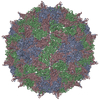
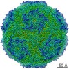
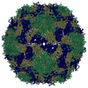
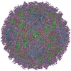
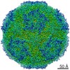
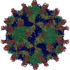
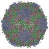
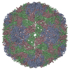
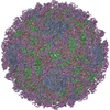
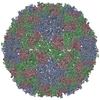
| Title | Structure and genome release mechanism of human cardiovirus Saffold virus-3 | ||||||
 Components Components |
| ||||||
 Keywords Keywords | VIRAL PROTEIN / SAFFOLD / VIRUS / CARDIOVIRUS / PICORNAVIRALES / A / ALTERED / VIRION / PARTICLE / CAPSID / GENOME / RNA / SSRNA | ||||||
| Function / homology |  Function and homology information Function and homology informationhost cell nucleolus / symbiont-mediated suppression of host mRNA export from nucleus / picornain 3C / T=pseudo3 icosahedral viral capsid / host cell cytoplasmic vesicle membrane / channel activity / monoatomic ion transmembrane transport / RNA helicase activity / RNA helicase / RNA-directed RNA polymerase ...host cell nucleolus / symbiont-mediated suppression of host mRNA export from nucleus / picornain 3C / T=pseudo3 icosahedral viral capsid / host cell cytoplasmic vesicle membrane / channel activity / monoatomic ion transmembrane transport / RNA helicase activity / RNA helicase / RNA-directed RNA polymerase / cysteine-type endopeptidase activity / viral RNA genome replication / RNA-directed RNA polymerase activity / symbiont entry into host cell / virion attachment to host cell / DNA-templated transcription / structural molecule activity / ATP hydrolysis activity / proteolysis / RNA binding / zinc ion binding / ATP binding Similarity search - Function | ||||||
| Biological species |  SAFFOLD VIRUS SAFFOLD VIRUS | ||||||
| Method | ELECTRON MICROSCOPY / single particle reconstruction / cryo EM / Resolution: 10.6 Å | ||||||
 Authors Authors | Mullapudi, E. / Novacek, J. / Palkova, L. / Kulich, P. / Lindberg, M. / vanKuppeveld, F.J.M. / Plevka, P. | ||||||
 Citation Citation | Journal: J Virol / Year: 2016 Title: Structure and Genome Release Mechanism of the Human Cardiovirus Saffold Virus 3. Authors: Edukondalu Mullapudi / Jiří Nováček / Lenka Pálková / Pavel Kulich / A Michael Lindberg / Frank J M van Kuppeveld / Pavel Plevka /    Abstract: In order to initiate an infection, viruses need to deliver their genomes into cells. This involves uncoating the genome and transporting it to the cytoplasm. The process of genome delivery is not ...In order to initiate an infection, viruses need to deliver their genomes into cells. This involves uncoating the genome and transporting it to the cytoplasm. The process of genome delivery is not well understood for nonenveloped viruses. We address this gap in our current knowledge by studying the uncoating of the nonenveloped human cardiovirus Saffold virus 3 (SAFV-3) of the family Picornaviridae SAFVs cause diseases ranging from gastrointestinal disorders to meningitis. We present a structure of a native SAFV-3 virion determined to 2.5 Å by X-ray crystallography and an 11-Å-resolution cryo-electron microscopy reconstruction of an "altered" particle that is primed for genome release. The altered particles are expanded relative to the native virus and contain pores in the capsid that might serve as channels for the release of VP4 subunits, N termini of VP1, and the RNA genome. Unlike in the related enteroviruses, pores in SAFV-3 are located roughly between the icosahedral 3- and 5-fold axes at an interface formed by two VP1 and one VP3 subunit. Furthermore, in native conditions many cardioviruses contain a disulfide bond formed by cysteines that are separated by just one residue. The disulfide bond is located in a surface loop of VP3. We determined the structure of the SAFV-3 virion in which the disulfide bonds are reduced. Disruption of the bond had minimal effect on the structure of the loop, but it increased the stability and decreased the infectivity of the virus. Therefore, compounds specifically disrupting or binding to the disulfide bond might limit SAFV infection. IMPORTANCE: A capsid assembled from viral proteins protects the virus genome during transmission from one cell to another. However, when a virus enters a cell the virus genome has to be released from ...IMPORTANCE: A capsid assembled from viral proteins protects the virus genome during transmission from one cell to another. However, when a virus enters a cell the virus genome has to be released from the capsid in order to initiate infection. This process is not well understood for nonenveloped viruses. We address this gap in our current knowledge by studying the genome release of Human Saffold virus 3 Saffold viruses cause diseases ranging from gastrointestinal disorders to meningitis. We show that before the genome is released, the Saffold virus 3 particle expands, and holes form in the previously compact capsid. These holes serve as channels for the release of the genome and small capsid proteins VP4 that in related enteroviruses facilitate subsequent transport of the virus genome into the cell cytoplasm. | ||||||
| History |
|
- Structure visualization
| Movie |
Movie viewer |
|---|---|
| Structure viewer | Molecule: MolmilJmol/JSmol |
- Downloads & links
-Download
| PDBx/mmCIF format | 5a8f.cif.gz | 141.8 KB | Display | PDBx/mmCIF format |
|---|---|---|---|---|
| PDB format | pdb5a8f.ent.gz | 109.7 KB | Display | PDB format |
| PDBx/mmJSON format | 5a8f.json.gz | Tree view | PDBx/mmJSON format | |
| Others |  Other downloads Other downloads |
-Validation report
| Arichive directory | https://data.pdbj.org/pub/pdb/validation_reports/a8/5a8fftp://data.pdbj.org/pub/pdb/validation_reports/a8/5a8f | HTTPS FTP |
|---|
-Related structure data
| Related structure data |  3097MC  5cfcC 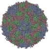 5cfdC M: map data used to model this data C: citing same article ( |
|---|---|
| Similar structure data |
-Links
 PDBj
PDBj

- Assembly
| Deposited unit | 
|
|---|---|
| 1 | x 60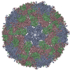
|
| 2 |
|
| 3 | x 5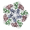
|
| 4 | x 6
|
| 5 | 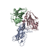
|
| Symmetry | Point symmetry: (Schoenflies symbol: I (icosahedral)) |
-Components
| #1: Protein | Mass: 24785.914 Da / Num. of mol.: 1 / Source method: isolated from a natural source / Source: (natural) SAFFOLD VIRUS / References: UniProt: C0MHL9 |
|---|---|
| #2: Protein | Mass: 20893.826 Da / Num. of mol.: 1 / Source method: isolated from a natural source / Source: (natural) SAFFOLD VIRUS / References: UniProt: E3TMG9 |
| #3: Protein | Mass: 28698.160 Da / Num. of mol.: 1 / Source method: isolated from a natural source / Source: (natural) SAFFOLD VIRUS / References: UniProt: C0MHL9 |
| Has protein modification | Y |
-Experimental details
-Experiment
| Experiment | Method: ELECTRON MICROSCOPY |
|---|---|
| EM experiment | Aggregation state: PARTICLE / 3D reconstruction method: single particle reconstruction |
- Sample preparation
| Component | Name: A PARTICLE OF SAFFOLD VIRUS-3 / Type: VIRUS |
|---|---|
| Buffer solution | Name: 20MM HEPES, 150MM NACL / pH: 7.5 / Details: 20MM HEPES, 150MM NACL |
| Specimen | Conc.: 2 mg/ml / Embedding applied: NO / Shadowing applied: NO / Staining applied: NO / Vitrification applied: YES |
| Specimen support | Details: HOLEY CARBON |
| Vitrification | Instrument: FEI VITROBOT MARK IV / Cryogen name: ETHANE Details: CRYOGEN - LIQUID ETHANE, VITROBOT MARK IV, BLOT FOR 2 SECONDS |
- Electron microscopy imaging
| Experimental equipment |  Model: Tecnai F20 / Image courtesy: FEI Company |
|---|---|
| Microscopy | Model: FEI TECNAI F20 / Date: Sep 3, 2014 |
| Electron gun | Electron source:  FIELD EMISSION GUN / Accelerating voltage: 200 kV / Illumination mode: SPOT SCAN FIELD EMISSION GUN / Accelerating voltage: 200 kV / Illumination mode: SPOT SCAN |
| Electron lens | Mode: BRIGHT FIELD / Nominal magnification: 55000 X / Nominal defocus max: 3950 nm / Nominal defocus min: 2330 nm / Cs: 2 mm |
| Specimen holder | Tilt angle max: 0 ° |
| Image recording | Electron dose: 20 e/Å2 / Film or detector model: FEI EAGLE (4k x 4k) |
| Image scans | Num. digital images: 264 |
- Processing
| EM software |
| ||||||||||||||||||||
|---|---|---|---|---|---|---|---|---|---|---|---|---|---|---|---|---|---|---|---|---|---|
| CTF correction | Details: EACH PARTICLE | ||||||||||||||||||||
| Symmetry | Point symmetry: I (icosahedral) | ||||||||||||||||||||
| 3D reconstruction | Method: PROJECTION MATCHING, FOURIER METHODS / Resolution: 10.6 Å / Num. of particles: 14028 / Nominal pixel size: 2.2176 Å / Actual pixel size: 2.2176 Å Details: SUBMISSION BASED ON EXPERIMENTAL DATA FROM EMDB EMD-3097. (DEPOSITION ID: 13604). Symmetry type: POINT | ||||||||||||||||||||
| Atomic model building | Protocol: OTHER / Space: REAL / Details: METHOD--LOCAL CORRELATION | ||||||||||||||||||||
| Refinement | Highest resolution: 10.6 Å | ||||||||||||||||||||
| Refinement step | Cycle: LAST / Highest resolution: 10.6 Å
|
