 Movie
Movie Controller

[English] 日本語
 Yorodumi
- PDB-1d3i: CRYO-EM STRUCTURE OF HUMAN RHINOVIRUS 14 (HRV14) COMPLEXED WITH A... -
+ Open data

- Basic information
| Entry | Database: PDB / ID: 1d3i | ||||||
|---|---|---|---|---|---|---|---|
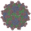
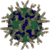
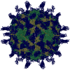
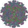
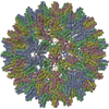
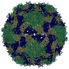
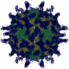
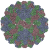
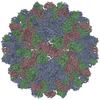
| Title | CRYO-EM STRUCTURE OF HUMAN RHINOVIRUS 14 (HRV14) COMPLEXED WITH A TWO-DOMAIN FRAGMENT OF ITS CELLULAR RECEPTOR, INTERCELLULAR ADHESION MOLECULE-1 (D1D2-ICAM-1). IMPLICATIONS FOR VIRUS-RECEPTOR INTERACTIONS. ALPHA CARBONS ONLY | ||||||
 Components Components |
| ||||||
 Keywords Keywords | Virus/Receptor / HUMAN RHINOVIRUS / HRV14 / ICAM-1 / FITTING OF X-RAY STRUCTURES INTO CRYO-EM RECONSTRUCTIONS / COMMON COLD / VIRUS UNCOATING / VIRUS/ VIRAL PROTEIN / RHINOVIRUS-RECEPTOR COMPLEX / Icosahedral virus / Virus-Receptor COMPLEX | ||||||
| Function / homology |  Function and homology information Function and homology informationregulation of leukocyte mediated cytotoxicity / T cell extravasation / positive regulation of cellular extravasation / leukocyte adhesion to vascular endothelial cell / regulation of ruffle assembly / membrane to membrane docking / T cell antigen processing and presentation / cell-cell adhesion mediated by integrin / T cell activation via T cell receptor contact with antigen bound to MHC molecule on antigen presenting cell / lysis of host organelle involved in viral entry into host cell ...regulation of leukocyte mediated cytotoxicity / T cell extravasation / positive regulation of cellular extravasation / leukocyte adhesion to vascular endothelial cell / regulation of ruffle assembly / membrane to membrane docking / T cell antigen processing and presentation / cell-cell adhesion mediated by integrin / T cell activation via T cell receptor contact with antigen bound to MHC molecule on antigen presenting cell / lysis of host organelle involved in viral entry into host cell / adhesion of symbiont to host / establishment of endothelial barrier / heterophilic cell-cell adhesion / leukocyte migration / leukocyte cell-cell adhesion / Interleukin-10 signaling / symbiont-mediated suppression of host cytoplasmic pattern recognition receptor signaling pathway via inhibition of RIG-I activity / immunological synapse / Integrin cell surface interactions / negative regulation of endothelial cell apoptotic process / negative regulation of extrinsic apoptotic signaling pathway via death domain receptors / cellular response to leukemia inhibitory factor / picornain 2A / symbiont-mediated suppression of host mRNA export from nucleus / symbiont genome entry into host cell via pore formation in plasma membrane / picornain 3C / cellular response to glucose stimulus / T=pseudo3 icosahedral viral capsid / host cell cytoplasmic vesicle membrane / integrin binding / cellular response to amyloid-beta / Interferon gamma signaling / Immunoregulatory interactions between a Lymphoid and a non-Lymphoid cell / ribonucleoside triphosphate phosphatase activity / transmembrane signaling receptor activity / nucleoside-triphosphate phosphatase / virus receptor activity / extracellular matrix / channel activity / signaling receptor activity / Interleukin-4 and Interleukin-13 signaling / monoatomic ion transmembrane transport / receptor-mediated virion attachment to host cell / positive regulation of ERK1 and ERK2 cascade / DNA replication / cell adhesion / RNA helicase activity / endocytosis involved in viral entry into host cell / membrane raft / receptor ligand activity / external side of plasma membrane / symbiont-mediated activation of host autophagy / RNA-directed RNA polymerase / cysteine-type endopeptidase activity / focal adhesion / viral RNA genome replication / RNA-directed RNA polymerase activity / virion attachment to host cell / host cell nucleus / DNA-templated transcription / structural molecule activity / cell surface / proteolysis / : / RNA binding / extracellular exosome / zinc ion binding / ATP binding / membrane / plasma membrane Similarity search - Function | ||||||
| Biological species |  Homo sapiens (human) Homo sapiens (human) Human rhinovirus sp. Human rhinovirus sp. | ||||||
| Method | ELECTRON MICROSCOPY / single particle reconstruction / cryo EM / Resolution: 26 Å | ||||||
 Authors Authors | Bella, J. / Rossmann, M.G. | ||||||
 Citation Citation | Journal: EMBO J / Year: 1999 Title: Structural studies of two rhinovirus serotypes complexed with fragments of their cellular receptor. Authors: P R Kolatkar / J Bella / N H Olson / C M Bator / T S Baker / M G Rossmann /  Abstract: Two human rhinovirus serotypes complexed with two- and five-domain soluble fragments of the cellular receptor, intercellular adhesion molecule-1, have been investigated by X-ray crystallographic ...Two human rhinovirus serotypes complexed with two- and five-domain soluble fragments of the cellular receptor, intercellular adhesion molecule-1, have been investigated by X-ray crystallographic analyses of the individual components and by cryo-electron microscopy of the complexes. The three-dimensional image reconstructions provide a molecular envelope within which the crystal structures of the viruses and the receptor fragments can be positioned with accuracy. The N-terminal domain of the receptor binds to the rhinovirus 'canyon' surrounding the icosahedral 5-fold axes. Fitting of molecular models into the image reconstruction density identified the residues on the virus that interact with those on the receptor surface, demonstrating complementarity of the electrostatic patterns for the tip of the N-terminal receptor domain and the floor of the canyon. The complexes seen in the image reconstructions probably represent the first stage of a multistep binding process. A mechanism is proposed for the subsequent viral uncoating process. #1: Journal: Proc.Natl.Acad.Sci.USA / Year: 1998Title: The Structure of the Two Amino-Terminal Domains of Human Icam-1 Suggests How It Functions as a Rhinovirus Receptor and as an Lfa-1 Integrin Ligand. Authors: Bella, J. / Kolatkar, P.R. / Marlor, C.W. / Greve, J.M. / Rossmann, M.G. #2: Journal: J.Mol.Biol. / Year: 1990Title: Analysis of the Structure of a Common Cold Virus, Human Rhinovirus 14, Refined at a Resolution of 3.0 Angstroms. Authors: Arnold, E. / Rossmann, M.G. #3: Journal: Nature / Year: 1985Title: Structure of a Human Common Cold Virus and Functional Relationship to Other Picornaviruses Authors: Rossmann, M.G. / Arnold, E. / Erickson, J.W. / Frankenberger, E.A. / Griffith, J.P. / Hecht, H.-J. / Johnson, J.E. / Kamer, G. / Luo, M. / Mosser, A.G. / Rueckert, R.R. / Sherry, B. / Vriend, G. #4: Journal: Proc.Natl.Acad.Sci.USA / Year: 1993Title: Structure of a Human Rhinovirus Complexed with its Receptor Molecule Authors: Olson, N.H. / Kolatkar, P.R. / Oliveira, M.A. / Cheng, R.H. / Greve, J.M. / Mcclelland, A. / Baker, T.S. / Rossmann, M.G. #5: Journal: Proc.Natl.Acad.Sci.USA / Year: 1998Title: A Dimeric Crystal Structure for the N-Terminal Two Domains of Intercellular Adhesion Molecule-1 Authors: Casasnovas, J.M. / Stehle, T. / Liu, J.H. / Wang, J.H. / Springer, T.A. | ||||||
| History |
|
- Structure visualization
| Movie |
Movie viewer |
|---|---|
| Structure viewer | Molecule: MolmilJmol/JSmol |
- Downloads & links
-Download
| PDBx/mmCIF format | 1d3i.cif.gz | 49 KB | Display | PDBx/mmCIF format |
|---|---|---|---|---|
| PDB format | pdb1d3i.ent.gz | 26.2 KB | Display | PDB format |
| PDBx/mmJSON format | 1d3i.json.gz | Tree view | PDBx/mmJSON format | |
| Others |  Other downloads Other downloads |
-Validation report
| Arichive directory | https://data.pdbj.org/pub/pdb/validation_reports/d3/1d3iftp://data.pdbj.org/pub/pdb/validation_reports/d3/1d3i | HTTPS FTP |
|---|
-Related structure data


-Links
 PDBj
PDBj


- Assembly
| Deposited unit | 
|
|---|---|
| 1 | x 60
|
| 2 |
|
| 3 | x 5
|
| 4 | x 6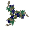
|
| 5 | 
|
| Symmetry | Point symmetry: (Hermann–Mauguin notation: 532 / Schoenflies symbol: I (icosahedral)) |
-Components
| #1: Protein | Mass: 20438.260 Da / Num. of mol.: 1 / Fragment: FIRST TWO DOMAINS, RESIDUES 1-185 / Source method: isolated from a natural source / Source: (natural) Homo sapiens (human) / Fragment: 1 - 185 / References: UniProt: P05362 |
|---|---|
| #2: Protein | Mass: 32560.549 Da / Num. of mol.: 1 / Source method: isolated from a natural source / Source: (natural) Human rhinovirus sp. / Genus: Rhinovirus / Strain: SEROTYPE 14 / References: UniProt: P03303 |
| #3: Protein | Mass: 28501.361 Da / Num. of mol.: 1 / Source method: isolated from a natural source / Source: (natural) Human rhinovirus sp. / Genus: Rhinovirus / Strain: SEROTYPE 14 / References: UniProt: P03303 |
| #4: Protein | Mass: 26236.754 Da / Num. of mol.: 1 / Source method: isolated from a natural source / Source: (natural) Human rhinovirus sp. / Genus: Rhinovirus / Strain: SEROTYPE 14 / References: UniProt: P03303 |
| #5: Protein | Mass: 7183.863 Da / Num. of mol.: 1 / Source method: isolated from a natural source / Source: (natural) Human rhinovirus sp. / Genus: Rhinovirus / Strain: SEROTYPE 14 / References: UniProt: P03303 |
-Experimental details
-Experiment
| Experiment | Method: ELECTRON MICROSCOPY |
|---|---|
| EM experiment | Aggregation state: PARTICLE / 3D reconstruction method: single particle reconstruction |
- Sample preparation
| Component | Name: HUMAN RHINOVIRUS 14 COMPLEXED WITH INTERCELLULAR ADHESION MOLECULE-1 Type: COMPLEX | ||||||||||||||||||||||||||||||||||||||||||
|---|---|---|---|---|---|---|---|---|---|---|---|---|---|---|---|---|---|---|---|---|---|---|---|---|---|---|---|---|---|---|---|---|---|---|---|---|---|---|---|---|---|---|---|
| Buffer solution | pH: 7.5 | ||||||||||||||||||||||||||||||||||||||||||
| Specimen | Embedding applied: NO / Shadowing applied: NO / Staining applied: NO / Vitrification applied: YES | ||||||||||||||||||||||||||||||||||||||||||
| Vitrification | Details: HRV14 WAS INCUBATED WITH D1D2-ICAM-1 FOR 30 MINUTES AT 4 DEGREES CELSIUS (277 KELVIN) USING AN EIGHT-FOLD EXCESS OF D1D2-ICAM-1 FOR EACH OF THE SIXTY POSSIBLE BINDING SITES PER VIRION. AFTER ...Details: HRV14 WAS INCUBATED WITH D1D2-ICAM-1 FOR 30 MINUTES AT 4 DEGREES CELSIUS (277 KELVIN) USING AN EIGHT-FOLD EXCESS OF D1D2-ICAM-1 FOR EACH OF THE SIXTY POSSIBLE BINDING SITES PER VIRION. AFTER INCUBATION, SAMPLES WERE PREPARED AS THIN LAYERS OF VITREOUS ICE AND MAINTAINED AT NEAR LIQUID NITROGEN TEMPERATURE IN THE ELECTRON MICROSCOPE WITH A GATAN 626 CRYOTRANSFER HOLDER. | ||||||||||||||||||||||||||||||||||||||||||
| Crystal grow | *PLUS Method: vapor diffusion, hanging drop | ||||||||||||||||||||||||||||||||||||||||||
| Components of the solutions | *PLUS
|
- Electron microscopy imaging
| Microscopy | Model: FEI/PHILIPS EM420 / Date: Jun 1, 1993 |
|---|---|
| Electron gun | Accelerating voltage: 80 kV / Illumination mode: FLOOD BEAM |
| Electron lens | Mode: BRIGHT FIELD / Nominal magnification: 49000 X / Nominal defocus max: 1250 nm |
| Specimen holder | Temperature: 120 K |
| Image recording | Electron dose: 20 e/Å2 / Film or detector model: KODAK SO-163 FILM |
- Processing
| EM software |
| ||||||||||||
|---|---|---|---|---|---|---|---|---|---|---|---|---|---|
| Symmetry | Point symmetry: I (icosahedral) | ||||||||||||
| 3D reconstruction | Method: MODEL-BASED, POLAR-FOURIER-TRANSFORM (FULLER ET AL. 1996, J.STRUC.BIOL.116, 48-55; BAKER AND CHENG, 1996, J.STRUC.BIOL. 116, 120-130) Resolution: 26 Å / Resolution method: OTHER / Num. of particles: 36 / Nominal pixel size: 5.1 Å / Actual pixel size: 5.19 Å Magnification calibration: THE PIXEL SIZE OF THE CRYO-EM MAP WAS CALIBRATED AGAINST THE CRYO-EM RECONSTRUCTION OF THE D1D2-ICAM-1/HRV16 COMPLEX. DENSITIES WERE COMPARED BY CROSS-CORRELATION WITHIN A ...Magnification calibration: THE PIXEL SIZE OF THE CRYO-EM MAP WAS CALIBRATED AGAINST THE CRYO-EM RECONSTRUCTION OF THE D1D2-ICAM-1/HRV16 COMPLEX. DENSITIES WERE COMPARED BY CROSS-CORRELATION WITHIN A SPHERICAL SHELL OF INTERNAL RADIUS 110 ANGSTROMS AND EXTERNAL RADIUS 216 ANGSTROMS. Details: THE RESOLUTION OF THE FINAL RECONSTRUCTED DENSITY WAS DETERMINED TO BE AT LEAST 26 ANGSTROMS, AS MEASURED BY RANDOMLY SPLITTING THE PARTICLES INTO TWO SETS AND COMPARING STRUCTURE FACTORS ...Details: THE RESOLUTION OF THE FINAL RECONSTRUCTED DENSITY WAS DETERMINED TO BE AT LEAST 26 ANGSTROMS, AS MEASURED BY RANDOMLY SPLITTING THE PARTICLES INTO TWO SETS AND COMPARING STRUCTURE FACTORS OBTAINED FROM SEPARATE RECONSTRUCTIONS (BAKER ET AL. 1991, BIOPHYS.J. 60, 1445-1456). THE EIGENVALUE SPECTRUM GAVE AN INDICATION OF THE RANDOMNESS OF THE DATA THAT WAS INCLUDED IN THE RECONSTRUCTION. THE COMPLETENESS OF THE DATA WAS VERIFIED IN THAT ALL EIGENVALUES EXCEEDED 1.0. Symmetry type: POINT | ||||||||||||
| Atomic model building | Protocol: RIGID BODY FIT / Space: RECIPROCAL / Target criteria: VECTOR R-FACTOR Details: REFINEMENT PROTOCOL--RIGID BODY REFINEMENT DETAILS--THE CRYSTAL STRUCTURE OF HRV14 WAS PLACED INTO THE CALIBRATED CRYO-EM DENSITY MAP BY ALIGNING THE ICOSAHEDRAL SYMMETRY AXES. APPROPRIATELY ...Details: REFINEMENT PROTOCOL--RIGID BODY REFINEMENT DETAILS--THE CRYSTAL STRUCTURE OF HRV14 WAS PLACED INTO THE CALIBRATED CRYO-EM DENSITY MAP BY ALIGNING THE ICOSAHEDRAL SYMMETRY AXES. APPROPRIATELY GLYCOSYLATED MODELS OF D1D2-ICAM-1 WITH VARIOUS INTERDOMAIN ANGLES (AS SEEN IN DIFFERENT CRYSTAL STRUCTURES OF D1D2-ICAM-1), WERE FIRST MANUALLY FITTED INTO THE CRYO-EM DENSITY CORRESPONDING TO THE ICAM-1 FRAGMENT, AND SUBSEQUENTLY REFINED AS RIGID BODIES IN RECIPROCAL SPACE. OBSERVED STRUCTURE FACTORS WERE OBTAINED BY INVERSE FOURIER TRANSFORM OF CRYO-EM DIFFERENCE MAPS CALCULATED BY 1) SUBSTRACTION OF THE HRV14 AND RNA CONTRIBUTION FROM THE CRYO-EM RECONSTRUCTED DENSITY OF THE COMPLEXES; 2) REDUCTION OF THE DIFFERENCE MAPS TO AN ICOSAHEDRAL ASYMMETRIC UNIT. THE COORDINATES ARE IN THE P, Q, R FRAME IN ANGSTROM UNITS AND CORRESPOND TO ICOSAHEDRAL SYMMETRY AXES. THE ORIGIN IS CHOSEN AT THE CENTER OF THE VIRUS WITH P, Q AND R ALONG MUTUALLY PERPENDICULAR TWO-FOLD AXES OF THE ICOSAHEDRON. THEY SHOULD REMAIN IN THAT FRAME FOR THE EASE OF THE USER IN CREATING THE BIOLOGICALLY SIGNIFICANT VIRAL COMPLEX PARTICLE USING THE 60 ICOSAHEDRAL SYMMETRY OPERATORS. RESIDUES NOT VISIBLE IN THE ORIGINAL CRYSTAL STRUCTURES ARE NOT INCLUDED IN THE CRYO-EM STRUCTURE MODEL. FOR EXAMPLE, HRV14 RESIDUES 1001-1016, 2001-2007 AND 4001-4028 ARE NOT VISIBLE IN THE CRYSTAL STRUCTURE (PDB ENTRY 4RHV) AND THEREFORE ARE NOT INCLUDED IN THE COORDINATES BELOW. | ||||||||||||
| Refinement | Highest resolution: 26 Å | ||||||||||||
| Refinement step | Cycle: LAST / Highest resolution: 26 Å
|
