 Movie
Movie Controller


+ Open data

- Basic information
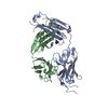
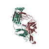
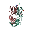
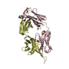
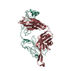
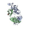
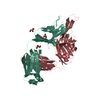
| Entry | Database: PDB / ID: 3eot | ||||||
|---|---|---|---|---|---|---|---|
| Title | Crystal structure of LAC031, an engineered anti-VLA1 Fab | ||||||
 Components Components |
| ||||||
 Keywords Keywords | IMMUNE SYSTEM / antibody / Fab / VLA-1 / domain swap / proten engineering | ||||||
| Function / homology | Immunoglobulins / Immunoglobulin-like / Sandwich / Mainly Beta Function and homology information Function and homology information | ||||||
| Biological species |  | ||||||
| Method |  X-RAY DIFFRACTION / SYNCHROTRON / Resolution: 1.9 Å X-RAY DIFFRACTION / SYNCHROTRON / Resolution: 1.9 Å | ||||||
 Authors Authors | Boriack-Sjodin, P.A. / Clark, L.A. | ||||||
 Citation Citation | Journal: Protein Eng.Des.Sel. / Year: 2009 Title: An antibody loop replacement design feasibility study and a loop-swapped dimer structure. Authors: Clark, L.A. / Boriack-Sjodin, P.A. / Day, E. / Eldredge, J. / Fitch, C. / Jarpe, M. / Miller, S. / Li, Y. / Simon, K. / van Vlijmen, H.W. | ||||||
| History |
|
- Structure visualization
| Structure viewer | Molecule: MolmilJmol/JSmol |
|---|
- Downloads & links
-Download
| PDBx/mmCIF format | 3eot.cif.gz | 97.3 KB | Display | PDBx/mmCIF format |
|---|---|---|---|---|
| PDB format | pdb3eot.ent.gz | 73.5 KB | Display | PDB format |
| PDBx/mmJSON format | 3eot.json.gz | Tree view | PDBx/mmJSON format | |
| Others |  Other downloads Other downloads |
-Validation report
| Summary document | 3eot_validation.pdf.gz | 436.7 KB | Display | wwPDB validaton report |
|---|---|---|---|---|
| Full document | 3eot_full_validation.pdf.gz | 445.1 KB | Display | |
| Data in XML | 3eot_validation.xml.gz | 19.2 KB | Display | |
| Data in CIF | 3eot_validation.cif.gz | 27.3 KB | Display | |
| Arichive directory | https://data.pdbj.org/pub/pdb/validation_reports/eo/3eotftp://data.pdbj.org/pub/pdb/validation_reports/eo/3eot | HTTPS FTP |
-Related structure data
| Related structure data | |
|---|---|
| Similar structure data |


-Links
 PDBj
PDBj

- Assembly
| Deposited unit | 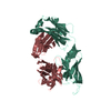
| ||||||||
|---|---|---|---|---|---|---|---|---|---|
| 1 |
| ||||||||
| Unit cell |
|
-Components
| #1: Antibody | Mass: 24224.074 Da / Num. of mol.: 1 / Mutation: W92I Source method: isolated from a genetically manipulated source Source: (gene. exp.)  |
|---|---|
| #2: Antibody | Mass: 23514.824 Da / Num. of mol.: 1 Source method: isolated from a genetically manipulated source Source: (gene. exp.) |
| #3: Water | ChemComp-HOH /  Mass: 18.015 Da / Num. of mol.: 204 / Source method: isolated from a natural source / Formula: H2O Mass: 18.015 Da / Num. of mol.: 204 / Source method: isolated from a natural source / Formula: H2O |
| Has protein modification | Y |
-Experimental details
-Experiment
| Experiment | Method: X-RAY DIFFRACTION / Number of used crystals: 1 |
|---|
- Sample preparation
| Crystal | Density Matthews: 2.41 Å3/Da / Density % sol: 49.01 % |
|---|---|
| Crystal grow | Temperature: 293 K / Method: vapor diffusion, hanging drop / pH: 6.5 Details: 16-20% Peg 3350, 100 mM Na Cacodylate, 200 mM MgCl2 , pH 6.5, VAPOR DIFFUSION, HANGING DROP, temperature 293K |
-Data collection
| Diffraction | Mean temperature: 100 K | ||||||||||||||||||||||||||||||||||||||||||||||||||||||||||||||||||
|---|---|---|---|---|---|---|---|---|---|---|---|---|---|---|---|---|---|---|---|---|---|---|---|---|---|---|---|---|---|---|---|---|---|---|---|---|---|---|---|---|---|---|---|---|---|---|---|---|---|---|---|---|---|---|---|---|---|---|---|---|---|---|---|---|---|---|---|
| Diffraction source | Source: SYNCHROTRON / Site: NSLS  / Beamline: X29A / Wavelength: 1.1 Å / Beamline: X29A / Wavelength: 1.1 Å | ||||||||||||||||||||||||||||||||||||||||||||||||||||||||||||||||||
| Detector | Type: ADSC QUANTUM 315 / Detector: CCD / Date: Mar 1, 2006 | ||||||||||||||||||||||||||||||||||||||||||||||||||||||||||||||||||
| Radiation | Protocol: SINGLE WAVELENGTH / Monochromatic (M) / Laue (L): M / Scattering type: x-ray | ||||||||||||||||||||||||||||||||||||||||||||||||||||||||||||||||||
| Radiation wavelength | Wavelength: 1.1 Å / Relative weight: 1 | ||||||||||||||||||||||||||||||||||||||||||||||||||||||||||||||||||
| Reflection | Resolution: 1.9→50 Å / Num. obs: 36217 / % possible obs: 96.3 % / Redundancy: 6.7 % / Rmerge(I) obs: 0.06 / Χ2: 1.169 / Net I/σ(I): 35.365 | ||||||||||||||||||||||||||||||||||||||||||||||||||||||||||||||||||
| Reflection shell |
|
- Processing
| Software |
| ||||||||||||||||||||||||||||||||||||
|---|---|---|---|---|---|---|---|---|---|---|---|---|---|---|---|---|---|---|---|---|---|---|---|---|---|---|---|---|---|---|---|---|---|---|---|---|---|
| Refinement | Resolution: 1.9→35 Å / Occupancy max: 1 / Occupancy min: 1 / FOM work R set: 0.792 / Cross valid method: THROUGHOUT / σ(F): 0 / Stereochemistry target values: Engh & Huber
| ||||||||||||||||||||||||||||||||||||
| Displacement parameters | Biso max: 66.08 Å2 / Biso mean: 34.008 Å2 / Biso min: 14.28 Å2
| ||||||||||||||||||||||||||||||||||||
| Refine analyze |
| ||||||||||||||||||||||||||||||||||||
| Refinement step | Cycle: LAST / Resolution: 1.9→35 Å
| ||||||||||||||||||||||||||||||||||||
| Refine LS restraints |
| ||||||||||||||||||||||||||||||||||||
| Xplor file |
|
