 Movie
Movie Controller

[English] 日本語
 Yorodumi
- PDB-3el4: Crystal structure of inhibitor saquinavir (SQV) complexed with th... -
+ Open data

- Basic information
| Entry | Database: PDB / ID: 3el4 | ||||||
|---|---|---|---|---|---|---|---|
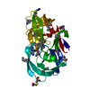
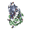
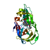
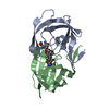
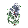
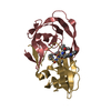
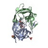
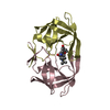
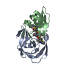
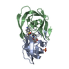
| Title | Crystal structure of inhibitor saquinavir (SQV) complexed with the multidrug HIV-1 protease variant L63P/V82T/I84V | ||||||
 Components Components | Protease | ||||||
 Keywords Keywords | HYDROLASE/HYDROLASE INHIBITOR / protease inhibitor / drug resistance / HIV protease / entropy-enthalpy compensation / AIDS / Hydrolase-Hydrolase inhibitor complex / Protease | ||||||
| Function / homology |  Function and homology information Function and homology informationHIV-1 retropepsin / symbiont-mediated activation of host apoptosis / retroviral ribonuclease H / exoribonuclease H / exoribonuclease H activity / DNA integration / viral genome integration into host DNA / establishment of integrated proviral latency / RNA-directed DNA polymerase / RNA stem-loop binding ...HIV-1 retropepsin / symbiont-mediated activation of host apoptosis / retroviral ribonuclease H / exoribonuclease H / exoribonuclease H activity / DNA integration / viral genome integration into host DNA / establishment of integrated proviral latency / RNA-directed DNA polymerase / RNA stem-loop binding / viral penetration into host nucleus / host multivesicular body / RNA-directed DNA polymerase activity / RNA-DNA hybrid ribonuclease activity / Transferases; Transferring phosphorus-containing groups; Nucleotidyltransferases / host cell / viral nucleocapsid / DNA recombination / DNA-directed DNA polymerase / aspartic-type endopeptidase activity / Hydrolases; Acting on ester bonds / DNA-directed DNA polymerase activity / symbiont-mediated suppression of host gene expression / viral translational frameshifting / symbiont entry into host cell / lipid binding / host cell nucleus / host cell plasma membrane / virion membrane / structural molecule activity / proteolysis / DNA binding / zinc ion binding Similarity search - Function | ||||||
| Biological species |  HIV-1 M:B_ARV2/SF2 (virus) HIV-1 M:B_ARV2/SF2 (virus) | ||||||
| Method |  X-RAY DIFFRACTION / MOLECULAR REPLACEMENT / molecular replacement / Resolution: 2 Å X-RAY DIFFRACTION / MOLECULAR REPLACEMENT / molecular replacement / Resolution: 2 Å | ||||||
 Authors Authors | Prabu-Jeyabalan, M. / King, N. / Schiffer, C. | ||||||
 Citation Citation | Journal: Acs Chem.Biol. / Year: 2012 Title: Extreme Entropy-Enthalpy Compensation in a Drug-Resistant Variant of HIV-1 Protease. Authors: King, N.M. / Prabu-Jeyabalan, M. / Bandaranayake, R.M. / Nalam, M.N. / Nalivaika, E.A. / Ozen, A. / Yilmaz, N.K. / Schiffer, C.A. | ||||||
| History |
|
- Structure visualization
| Structure viewer | Molecule: MolmilJmol/JSmol |
|---|
- Downloads & links
-Download
| PDBx/mmCIF format | 3el4.cif.gz | 54.7 KB | Display | PDBx/mmCIF format |
|---|---|---|---|---|
| PDB format | pdb3el4.ent.gz | 38.9 KB | Display | PDB format |
| PDBx/mmJSON format | 3el4.json.gz | Tree view | PDBx/mmJSON format | |
| Others |  Other downloads Other downloads |
-Validation report
| Arichive directory | https://data.pdbj.org/pub/pdb/validation_reports/el/3el4ftp://data.pdbj.org/pub/pdb/validation_reports/el/3el4 | HTTPS FTP |
|---|
-Related structure data
| Related structure data |  3ekpC  3ekqC  3ektC  3ekvC 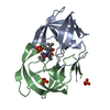 3ekwC  3ekxC  3ekyC  3el0C  3el1C  3el5C  3el9C 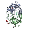 1f7aS C: citing same article ( S: Starting model for refinement |
|---|---|
| Similar structure data |


-Links
 PDBj
PDBj

- Assembly
| Deposited unit | 
| ||||||||
|---|---|---|---|---|---|---|---|---|---|
| 1 |
| ||||||||
| Unit cell |
| ||||||||
| Details | One dimer (A, B identifiers) and on saquinavir inhibitor in one asymmetric unit |
-Components
| #1: Protein | Mass: 10803.736 Da / Num. of mol.: 2 / Fragment: UNP residues 491-589 / Mutation: Q7K, V64I, V82T, I84V Source method: isolated from a genetically manipulated source Source: (gene. exp.) HIV-1 M:B_ARV2/SF2 (virus) / Strain: HXB2 / Gene: gag-pol / Plasmid: PXC35 / Production host:  #2: Chemical | ChemComp-ROC / ( |   Type: peptide-like, Peptide-like / Class: Inhibitor / Mass: 670.841 Da / Num. of mol.: 1 / Source method: obtained synthetically / Formula: C38H50N6O5 / Details: chemically synthesized / References: Saquinavir / Comment: medication, antiretroviral*YM Type: peptide-like, Peptide-like / Class: Inhibitor / Mass: 670.841 Da / Num. of mol.: 1 / Source method: obtained synthetically / Formula: C38H50N6O5 / Details: chemically synthesized / References: Saquinavir / Comment: medication, antiretroviral*YM#3: Chemical |   Mass: 59.044 Da / Num. of mol.: 2 / Source method: obtained synthetically / Formula: C2H3O2 Mass: 59.044 Da / Num. of mol.: 2 / Source method: obtained synthetically / Formula: C2H3O2#4: Water | ChemComp-HOH / |  Mass: 18.015 Da / Num. of mol.: 93 / Source method: isolated from a natural source / Formula: H2O Mass: 18.015 Da / Num. of mol.: 93 / Source method: isolated from a natural source / Formula: H2ONonpolymer details | THE INHIBITOR ROC IS A HYDROXYETH | |
|---|
-Experimental details
-Experiment
| Experiment | Method: X-RAY DIFFRACTION / Number of used crystals: 1 |
|---|
- Sample preparation
| Crystal | Density Matthews: 2.18 Å3/Da / Density % sol: 43.7 % |
|---|---|
| Crystal grow | Temperature: 300 K / Method: vapor diffusion, hanging drop / pH: 6.2 Details: pH 6.2, VAPOR DIFFUSION, HANGING DROP, temperature 300K |
-Data collection
| Diffraction | Mean temperature: 300 K | ||||||||||||||||||||||||||||||||||||||||||||||||||||||||||||||||||
|---|---|---|---|---|---|---|---|---|---|---|---|---|---|---|---|---|---|---|---|---|---|---|---|---|---|---|---|---|---|---|---|---|---|---|---|---|---|---|---|---|---|---|---|---|---|---|---|---|---|---|---|---|---|---|---|---|---|---|---|---|---|---|---|---|---|---|---|
| Diffraction source | Source: ROTATING ANODE / Type: RIGAKU / Wavelength: 1.5418 Å | ||||||||||||||||||||||||||||||||||||||||||||||||||||||||||||||||||
| Detector | Type: RIGAKU RAXIS IV / Detector: IMAGE PLATE / Details: Yale mirrors | ||||||||||||||||||||||||||||||||||||||||||||||||||||||||||||||||||
| Radiation | Monochromator: Yale Mirrors / Protocol: SINGLE WAVELENGTH / Monochromatic (M) / Laue (L): M / Scattering type: x-ray | ||||||||||||||||||||||||||||||||||||||||||||||||||||||||||||||||||
| Radiation wavelength | Wavelength: 1.5418 Å / Relative weight: 1 | ||||||||||||||||||||||||||||||||||||||||||||||||||||||||||||||||||
| Reflection | Resolution: 2→50 Å / Num. all: 12975 / Num. obs: 12975 / % possible obs: 97.2 % / Observed criterion σ(F): 0 / Observed criterion σ(I): 0 / Rmerge(I) obs: 0.099 / Χ2: 0.656 / Net I/σ(I): 14.091 | ||||||||||||||||||||||||||||||||||||||||||||||||||||||||||||||||||
| Reflection shell |
|
-Phasing
| Phasing | Method: molecular replacement |
|---|
- Processing
| Software |
| ||||||||||||||||||||||||
|---|---|---|---|---|---|---|---|---|---|---|---|---|---|---|---|---|---|---|---|---|---|---|---|---|---|
| Refinement | Method to determine structure: MOLECULAR REPLACEMENT Starting model: PDB ENTRY 1F7A Resolution: 2→50 Å / Occupancy max: 1 / Occupancy min: 0.5 / Isotropic thermal model: isotropic / Cross valid method: THROUGHOUT / σ(F): 0 / Stereochemistry target values: Engh & Huber
| ||||||||||||||||||||||||
| Solvent computation | Bsol: 57.411 Å2 | ||||||||||||||||||||||||
| Displacement parameters | Biso max: 57.29 Å2 / Biso mean: 25.534 Å2 / Biso min: 10.9 Å2
| ||||||||||||||||||||||||
| Refinement step | Cycle: LAST / Resolution: 2→50 Å
| ||||||||||||||||||||||||
| Refine LS restraints |
| ||||||||||||||||||||||||
| Xplor file |
|
