 Movie
Movie Controller

[English] 日本語
 Yorodumi
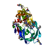
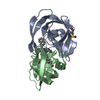
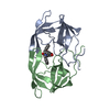
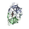
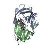
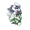
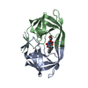
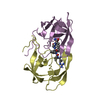
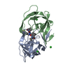
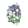
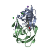
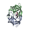
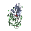
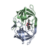
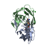
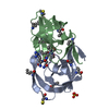
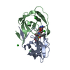
- PDB-3hlo: Crystal structure of chemically synthesized 'covalent dimer' [Gly... -
+ Open data

- Basic information
| Entry | Database: PDB / ID: 3hlo | ||||||
|---|---|---|---|---|---|---|---|
| Title | Crystal structure of chemically synthesized 'covalent dimer' [Gly51/D-Ala51']HIV-1 protease | ||||||
 Components Components | 'covalent dimer' [Gly51/D-Ala51'] HIV-1 protease | ||||||
 Keywords Keywords | HYDROLASE/HYDROLASE INHIBITOR / beta barrel / HYDROLASE / HYDROLASE-HYDROLASE INHIBITOR complex | ||||||
| Function / homology |  Function and homology information Function and homology informationHIV-1 retropepsin / symbiont-mediated activation of host apoptosis / retroviral ribonuclease H / exoribonuclease H / exoribonuclease H activity / DNA integration / viral genome integration into host DNA / establishment of integrated proviral latency / RNA-directed DNA polymerase / RNA stem-loop binding ...HIV-1 retropepsin / symbiont-mediated activation of host apoptosis / retroviral ribonuclease H / exoribonuclease H / exoribonuclease H activity / DNA integration / viral genome integration into host DNA / establishment of integrated proviral latency / RNA-directed DNA polymerase / RNA stem-loop binding / viral penetration into host nucleus / host multivesicular body / RNA-directed DNA polymerase activity / RNA-DNA hybrid ribonuclease activity / Transferases; Transferring phosphorus-containing groups; Nucleotidyltransferases / host cell / viral nucleocapsid / DNA recombination / DNA-directed DNA polymerase / aspartic-type endopeptidase activity / Hydrolases; Acting on ester bonds / DNA-directed DNA polymerase activity / symbiont-mediated suppression of host gene expression / viral translational frameshifting / symbiont entry into host cell / lipid binding / host cell nucleus / host cell plasma membrane / virion membrane / structural molecule activity / proteolysis / DNA binding / zinc ion binding Similarity search - Function | ||||||
| Method |  X-RAY DIFFRACTION / SYNCHROTRON / MOLECULAR REPLACEMENT / Resolution: 1.6 Å X-RAY DIFFRACTION / SYNCHROTRON / MOLECULAR REPLACEMENT / Resolution: 1.6 Å | ||||||
 Authors Authors | Torbeev, V.Y. / Kent, S.B.H. | ||||||
 Citation Citation | Journal: Proc.Natl.Acad.Sci.USA / Year: 2011 Title: Protein conformational dynamics in the mechanism of HIV-1 protease catalysis. Authors: Torbeev, V.Y. / Raghuraman, H. / Hamelberg, D. / Tonelli, M. / Westler, W.M. / Perozo, E. / Kent, S.B. | ||||||
| History |
|
- Structure visualization
| Structure viewer | Molecule: MolmilJmol/JSmol |
|---|
- Downloads & links
-Download
| PDBx/mmCIF format | 3hlo.cif.gz | 58 KB | Display | PDBx/mmCIF format |
|---|---|---|---|---|
| PDB format | pdb3hlo.ent.gz | 41.4 KB | Display | PDB format |
| PDBx/mmJSON format | 3hlo.json.gz | Tree view | PDBx/mmJSON format | |
| Others |  Other downloads Other downloads |
-Validation report
| Arichive directory | https://data.pdbj.org/pub/pdb/validation_reports/hl/3hloftp://data.pdbj.org/pub/pdb/validation_reports/hl/3hlo | HTTPS FTP |
|---|
-Related structure data
| Related structure data |  3fsmC  3hauC  3hawC  3hboC  3hdkC  3iawC  3ka2C  3nwqC  3nwxC 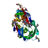 3nxeC  3nxnC  3nygC C: citing same article ( |
|---|---|
| Similar structure data |


-Links
 PDBj
PDBj

- Assembly
| Deposited unit | 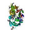
| ||||||||
|---|---|---|---|---|---|---|---|---|---|
| 1 |
| ||||||||
| Unit cell |
|
-Components
| #1: Protein | ' Mass: 21905.678 Da / Num. of mol.: 1 / Source method: obtained synthetically / Details: total chemical synthesis / References: UniProt: P03369*PLUS, HIV-1 retropepsin |
|---|---|
| #2: Chemical | ChemComp-2NC / 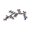  Type: peptide-like, Peptide-like / Class: Inhibitor / Mass: 770.983 Da / Num. of mol.: 1 / Source method: obtained synthetically / Formula: C35H68N11O8 Type: peptide-like, Peptide-like / Class: Inhibitor / Mass: 770.983 Da / Num. of mol.: 1 / Source method: obtained synthetically / Formula: C35H68N11O8References: N-{(2S)-2-[(N-acetyl-L-threonyl-L-isoleucyl)amino]hexyl}-L-norleucyl-L-glutaminyl-N~5~-[amino(iminio)methyl]-L-ornithinamide |
| #3: Water | ChemComp-HOH /  Mass: 18.015 Da / Num. of mol.: 141 / Source method: isolated from a natural source / Formula: H2O Mass: 18.015 Da / Num. of mol.: 141 / Source method: isolated from a natural source / Formula: H2O |
| Has protein modification | Y |
-Experimental details
-Experiment
| Experiment | Method: X-RAY DIFFRACTION / Number of used crystals: 1 |
|---|
- Sample preparation
| Crystal | Density Matthews: 2.09 Å3/Da / Density % sol: 41.08 % |
|---|---|
| Crystal grow | Temperature: 293 K / Method: vapor diffusion, hanging drop / pH: 6 Details: 0.1 M citrate, 0.2 M sodium phosphate, 30% (v/v) ammonium sulfate, 10% (v/v) DMSO, pH 6.0, VAPOR DIFFUSION, HANGING DROP, temperature 293K |
-Data collection
| Diffraction | Mean temperature: 110 K |
|---|---|
| Diffraction source | Source: SYNCHROTRON / Site: APS  / Beamline: 14-BM-C / Wavelength: 0.9002 Å / Beamline: 14-BM-C / Wavelength: 0.9002 Å |
| Detector | Type: ADSC QUANTUM 315 / Detector: CCD / Date: Feb 26, 2007 Details: bent conical Si-mirror (Rh coating); bent Ge(111) monochromator |
| Radiation | Monochromator: bent Ge(111) / Protocol: SINGLE WAVELENGTH / Monochromatic (M) / Laue (L): M / Scattering type: x-ray |
| Radiation wavelength | Wavelength: 0.9002 Å / Relative weight: 1 |
| Reflection | Resolution: 1.6→50 Å / Num. all: 25025 / Num. obs: 24925 / % possible obs: 99.6 % / Observed criterion σ(F): 0 / Observed criterion σ(I): 0 / Redundancy: 7.3 % / Rmerge(I) obs: 0.047 / Net I/σ(I): 38.3 |
| Reflection shell | Resolution: 1.6→1.66 Å / Redundancy: 7.1 % / Rmerge(I) obs: 0.354 / Mean I/σ(I) obs: 6.61 / Num. unique all: 2450 / % possible all: 100 |
- Processing
| Software |
| ||||||||||||||||||||||||||||||||||||||||||||||||||||||||||||||||||||||||||||||||||||||||||
|---|---|---|---|---|---|---|---|---|---|---|---|---|---|---|---|---|---|---|---|---|---|---|---|---|---|---|---|---|---|---|---|---|---|---|---|---|---|---|---|---|---|---|---|---|---|---|---|---|---|---|---|---|---|---|---|---|---|---|---|---|---|---|---|---|---|---|---|---|---|---|---|---|---|---|---|---|---|---|---|---|---|---|---|---|---|---|---|---|---|---|---|
| Refinement | Method to determine structure: MOLECULAR REPLACEMENT / Resolution: 1.6→20 Å / Cor.coef. Fo:Fc: 0.963 / Cor.coef. Fo:Fc free: 0.952 / SU B: 2.278 / SU ML: 0.062 / Cross valid method: THROUGHOUT / σ(F): 0 / σ(I): 0 / ESU R: 0.099 / ESU R Free: 0.098 / Stereochemistry target values: MAXIMUM LIKELIHOOD / Details: HYDROGENS HAVE BEEN ADDED IN THE RIDING POSITIONS
| ||||||||||||||||||||||||||||||||||||||||||||||||||||||||||||||||||||||||||||||||||||||||||
| Solvent computation | Ion probe radii: 0.8 Å / Shrinkage radii: 0.8 Å / VDW probe radii: 1.2 Å / Solvent model: BABINET MODEL WITH MASK | ||||||||||||||||||||||||||||||||||||||||||||||||||||||||||||||||||||||||||||||||||||||||||
| Displacement parameters | Biso mean: 25.329 Å2
| ||||||||||||||||||||||||||||||||||||||||||||||||||||||||||||||||||||||||||||||||||||||||||
| Refinement step | Cycle: LAST / Resolution: 1.6→20 Å
| ||||||||||||||||||||||||||||||||||||||||||||||||||||||||||||||||||||||||||||||||||||||||||
| Refine LS restraints |
| ||||||||||||||||||||||||||||||||||||||||||||||||||||||||||||||||||||||||||||||||||||||||||
| LS refinement shell | Resolution: 1.597→1.638 Å / Total num. of bins used: 20
|
