| Entry | Database: PDB / ID: 4k4p
|
|---|
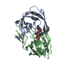
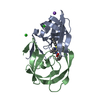
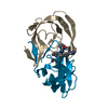
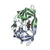
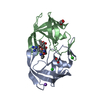
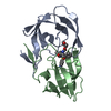
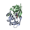
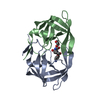
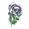
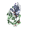
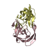
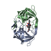
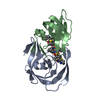
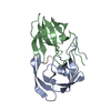
| Title | TL-3 inhibited Trp6Ala HIV Protease |
|---|
 Components Components | HIV-1 protease |
|---|
 Keywords Keywords | HYDROLASE/HYDROLASE INHIBITOR / fragment binding / exosite / protease / HYDROLASE-HYDROLASE INHIBITOR complex |
|---|
| Function / homology |  Function and homology information Function and homology information
HIV-1 retropepsin / symbiont-mediated activation of host apoptosis / retroviral ribonuclease H / exoribonuclease H / exoribonuclease H activity / DNA integration / viral genome integration into host DNA / establishment of integrated proviral latency / RNA-directed DNA polymerase / RNA stem-loop binding ...HIV-1 retropepsin / symbiont-mediated activation of host apoptosis / retroviral ribonuclease H / exoribonuclease H / exoribonuclease H activity / DNA integration / viral genome integration into host DNA / establishment of integrated proviral latency / RNA-directed DNA polymerase / RNA stem-loop binding / viral penetration into host nucleus / host multivesicular body / RNA-directed DNA polymerase activity / RNA-DNA hybrid ribonuclease activity / Transferases; Transferring phosphorus-containing groups; Nucleotidyltransferases / host cell / viral nucleocapsid / DNA recombination / DNA-directed DNA polymerase / aspartic-type endopeptidase activity / Hydrolases; Acting on ester bonds / DNA-directed DNA polymerase activity / symbiont-mediated suppression of host gene expression / viral translational frameshifting / symbiont entry into host cell / lipid binding / host cell nucleus / host cell plasma membrane / virion membrane / structural molecule activity / proteolysis / DNA binding / zinc ion bindingSimilarity search - Function Reverse transcriptase connection / Reverse transcriptase connection domain / Reverse transcriptase thumb / Reverse transcriptase thumb domain / Integrase Zinc binding domain / Zinc finger integrase-type profile. / Integrase DNA binding domain / Integrase, C-terminal domain superfamily, retroviral / Integrase, N-terminal zinc-binding domain / Integrase-like, N-terminal ...Reverse transcriptase connection / Reverse transcriptase connection domain / Reverse transcriptase thumb / Reverse transcriptase thumb domain / Integrase Zinc binding domain / Zinc finger integrase-type profile. / Integrase DNA binding domain / Integrase, C-terminal domain superfamily, retroviral / Integrase, N-terminal zinc-binding domain / Integrase-like, N-terminal / Integrase, C-terminal, retroviral / Integrase DNA binding domain profile. / Immunodeficiency lentiviral matrix, N-terminal / gag gene protein p17 (matrix protein) / RNase H / Integrase core domain / Integrase, catalytic core / Integrase catalytic domain profile. / Retropepsin-like catalytic domain / Matrix protein, lentiviral and alpha-retroviral, N-terminal / RNase H type-1 domain profile. / Ribonuclease H domain / Retroviral nucleocapsid Gag protein p24, C-terminal domain / Gag protein p24 C-terminal domain / Retropepsins / Retroviral aspartyl protease / Aspartyl protease, retroviral-type family profile. / Peptidase A2A, retrovirus, catalytic / Retrovirus capsid, C-terminal / Reverse transcriptase (RNA-dependent DNA polymerase) / Cathepsin D, subunit A; domain 1 / Acid Proteases / Retroviral matrix protein / Reverse transcriptase domain / Reverse transcriptase (RT) catalytic domain profile. / Retrovirus capsid, N-terminal / zinc finger / Zinc knuckle / Zinc finger, CCHC-type superfamily / Zinc finger, CCHC-type / Zinc finger CCHC-type profile. / Aspartic peptidase, active site / Eukaryotic and viral aspartyl proteases active site. / Aspartic peptidase domain superfamily / Ribonuclease H superfamily / Ribonuclease H-like superfamily / Reverse transcriptase/Diguanylate cyclase domain / DNA/RNA polymerase superfamily / Beta Barrel / Mainly BetaSimilarity search - Domain/homology N-[(benzyloxy)carbonyl]-L-alanyl-N-[(1R)-1-benzyl-2-oxoethyl]-L-valinamide / Chem-3TL / NITRATE ION / Gag-Pol polyproteinSimilarity search - Component |
|---|
| Biological species |   Human immunodeficiency virus type 1 Human immunodeficiency virus type 1 |
|---|
| Method |  X-RAY DIFFRACTION / SYNCHROTRON / MOLECULAR REPLACEMENT / molecular replacement / Resolution: 2.31 Å X-RAY DIFFRACTION / SYNCHROTRON / MOLECULAR REPLACEMENT / molecular replacement / Resolution: 2.31 Å |
|---|
 Authors Authors | Tiefenbrunn, T. / Stout, C.D. |
|---|
 Citation Citation | Journal: Chem.Biol.Drug Des. / Year: 2014
Title: Crystallographic Fragment-Based Drug Discovery: Use of a Brominated Fragment Library Targeting HIV Protease.
Authors: Tiefenbrunn, T. / Forli, S. / Happer, M. / Gonzalez, A. / Tsai, Y. / Soltis, M. / Elder, J.H. / Olson, A.J. / Stout, C.D. |
|---|
| History | | Deposition | Apr 12, 2013 | Deposition site: RCSB / Processing site: RCSB |
|---|
| Revision 1.0 | Sep 18, 2013 | Provider: repository / Type: Initial release |
|---|
| Revision 1.1 | Oct 16, 2013 | Group: Non-polymer description |
|---|
| Revision 1.2 | Feb 12, 2014 | Group: Database references |
|---|
| Revision 1.3 | Feb 28, 2024 | Group: Data collection / Database references / Derived calculations
Category: chem_comp_atom / chem_comp_bond ...chem_comp_atom / chem_comp_bond / database_2 / struct_ref_seq_dif / struct_site
Item: _database_2.pdbx_DOI / _database_2.pdbx_database_accession ..._database_2.pdbx_DOI / _database_2.pdbx_database_accession / _struct_ref_seq_dif.details / _struct_site.pdbx_auth_asym_id / _struct_site.pdbx_auth_comp_id / _struct_site.pdbx_auth_seq_id |
|---|
|
|---|
 Movie
Movie Controller


 Open data
 Basic information
 Structure visualization
 Downloads & links
 Other downloads


 PDBj
PDBj


 Assembly


 Mass: 62.005 Da / Num. of mol.: 1 / Source method: obtained synthetically / Formula: NO3
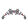
 Type: peptide-like, Peptide-like / Class: Inhibitor / Mass: 909.077 Da / Num. of mol.: 1 / Source method: obtained synthetically / Formula: C50H64N6O10
 Mass: 18.015 Da / Num. of mol.: 20 / Source method: isolated from a natural source / Formula: H2O
 Sample preparation
 / Beamline: BL11-1 / Wavelength: 0.918 Å
 Processing