- PDB-3n3i: Crystal Structure of G48V/C95F tethered HIV-1 Protease/Saquinavir... -
+
Open data
ID or keywords:
Loading...
-
Basic information
Entry
Database: PDB / ID: 3n3i
Title
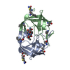
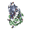
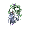
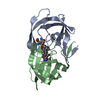
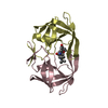
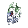
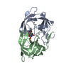
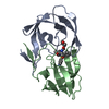
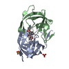
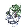
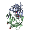
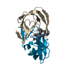
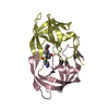
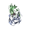
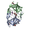
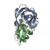
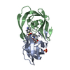
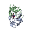
Crystal Structure of G48V/C95F tethered HIV-1 Protease/Saquinavir complex
Components
Protease
Keywords
HYDROLASE/HYDROLASE INHIBITOR / HIV-1 protease / Saquinavir / Drug Resistance / tethered-dimer / fusion protein / HYDROLASE-HYDROLASE INHIBITOR COMPLEX
Function / homology
Function and homology information
integrase activity / Integration of viral DNA into host genomic DNA / Autointegration results in viral DNA circles / Minus-strand DNA synthesis / Plus-strand DNA synthesis / Uncoating of the HIV Virion / 2-LTR circle formation / Vpr-mediated nuclear import of PICs / Early Phase of HIV Life Cycle / Integration of provirus ...integrase activity / Integration of viral DNA into host genomic DNA / Autointegration results in viral DNA circles / Minus-strand DNA synthesis / Plus-strand DNA synthesis / Uncoating of the HIV Virion / 2-LTR circle formation / Vpr-mediated nuclear import of PICs / Early Phase of HIV Life Cycle / Integration of provirus / APOBEC3G mediated resistance to HIV-1 infection / Binding and entry of HIV virion / viral life cycle / HIV-1 retropepsin / symbiont-mediated activation of host apoptosis / retroviral ribonuclease H / exoribonuclease H / exoribonuclease H activity / Assembly Of The HIV Virion / protein processing / viral genome integration into host DNA / Budding and maturation of HIV virion / establishment of integrated proviral latency / RNA-directed DNA polymerase / RNA stem-loop binding / viral penetration into host nucleus / host multivesicular body / RNA-directed DNA polymerase activity / RNA-DNA hybrid ribonuclease activity / Transferases; Transferring phosphorus-containing groups; Nucleotidyltransferases / peptidase activity / host cell / viral nucleocapsid / DNA recombination / DNA-directed DNA polymerase / aspartic-type endopeptidase activity / Hydrolases; Acting on ester bonds / DNA-directed DNA polymerase activity / symbiont-mediated suppression of host gene expression / viral translational frameshifting / symbiont entry into host cell / lipid binding / host cell nucleus / host cell plasma membrane / virion membrane / structural molecule activity / DNA binding / zinc ion binding / identical protein binding Similarity search - Function
Mass: 22078.994 Da / Num. of mol.: 1 / Fragment: UNP residues 489-587 / Mutation: G48V, G1048V, C95F, C1095F Source method: isolated from a genetically manipulated source Details: Chimera protein with LINKER Gly-Gly-Ser-Ser-Gly (numbered A100 to A104) that covalently link sub-unit A with sub-unit B in the tethered dimer. Source: (gene. exp.) Human immunodeficiency virus type 1 / Strain: group M subtype B (isolate HXB2) / Gene: gag-pol / Plasmid: pET11A / Production host: Escherichia coli (E. coli) / Strain (production host): BL21(DE3) / References: UniProt: P04585, HIV-1 retropepsin
Mass: 18.015 Da / Num. of mol.: 60 / Source method: isolated from a natural source / Formula: H2O
Nonpolymer details
THE INHIBITOR SAQUINAVIR (ROC) IS A HYDROXYETHYLAMINE CONTAINING TRANSITION STATE MIMETIC. IT IS ...THE INHIBITOR SAQUINAVIR (ROC) IS A HYDROXYETHYLAMINE CONTAINING TRANSITION STATE MIMETIC. IT IS REFERRED AS SQV IN THE ARTICLE.
Sequence details
THE LINKER IS DISORDERED. SO THERE ARE NO COORDINATES FOR THE LINKER GGSSG.
-
Experimental details
-
Experiment
Experiment
Method: X-RAY DIFFRACTION / Number of used crystals: 1
-
Sample preparation
Crystal
Density Matthews: 2.12 Å3/Da / Density % sol: 41.98 %
Crystal grow
Temperature: 298 K / Method: vapor diffusion, hanging drop / pH: 6.2 Details: 1-5% saturated Ammonium Sulfate, 200/100mM Phosphate/Citrate Buffer, pH 6.2, VAPOR DIFFUSION, HANGING DROP, temperature 298K
In the structure databanks used in Yorodumi, some data are registered as the other names, "COVID-19 virus" and "2019-nCoV". Here are the details of the virus and the list of structure data.
Jan 31, 2019. EMDB accession codes are about to change! (news from PDBe EMDB page)
EMDB accession codes are about to change! (news from PDBe EMDB page)
The allocation of 4 digits for EMDB accession codes will soon come to an end. Whilst these codes will remain in use, new EMDB accession codes will include an additional digit and will expand incrementally as the available range of codes is exhausted. The current 4-digit format prefixed with “EMD-” (i.e. EMD-XXXX) will advance to a 5-digit format (i.e. EMD-XXXXX), and so on. It is currently estimated that the 4-digit codes will be depleted around Spring 2019, at which point the 5-digit format will come into force.
The EM Navigator/Yorodumi systems omit the EMD- prefix.
Related info.:Q: What is EMD? / ID/Accession-code notation in Yorodumi/EM Navigator
Yorodumi is a browser for structure data from EMDB, PDB, SASBDB, etc.
This page is also the successor to EM Navigator detail page, and also detail information page/front-end page for Omokage search.
The word "yorodu" (or yorozu) is an old Japanese word meaning "ten thousand". "mi" (miru) is to see.
Related info.:EMDB / PDB / SASBDB / Comparison of 3 databanks / Yorodumi Search / Aug 31, 2016. New EM Navigator & Yorodumi / Yorodumi Papers / Jmol/JSmol / Function and homology information / Changes in new EM Navigator and Yorodumi
 Movie
Movie Controller

 Yorodumi
 Open data
 Basic information
 Components
 Keywords
 Function and homology information
 Human immunodeficiency virus type 1
 X-RAY DIFFRACTION /
  Authors
 Citation
 Structure visualization
 Downloads & links
 Other downloads
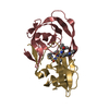

 PDBj
PDBj


 Assembly
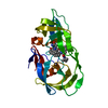


 Type: peptide-like, Peptide-like / Class: Inhibitor / Mass: 670.841 Da / Num. of mol.: 1 / Source method: obtained synthetically / Formula: C38H50N6O5 / References: Saquinavir
 Mass: 18.015 Da / Num. of mol.: 60 / Source method: isolated from a natural source / Formula: H2O
 Sample preparation
 / Beamline: X06DA / Wavelength: 1 Å
 Processing