 Movie
Movie Controller

[English] 日本語
 Yorodumi
- PDB-3eac: Crystal structure of SH2 domain of Human Csk (carboxyl-terminal s... -
+ Open data

- Basic information
| Entry | Database: PDB / ID: 3eac | ||||||
|---|---|---|---|---|---|---|---|
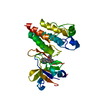
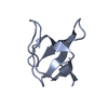
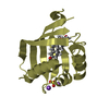
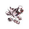
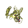
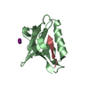
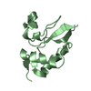
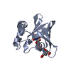
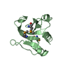
| Title | Crystal structure of SH2 domain of Human Csk (carboxyl-terminal src kinase), Oxidized form. | ||||||
 Components Components | Tyrosine-protein kinase CSK | ||||||
 Keywords Keywords | TRANSFERASE / SH2 / CSK / DISULFIDE / OXIDIZED / Reduced / ATP-binding / Cell membrane / Kinase / Membrane / Nucleotide-binding / Phosphoprotein / SH2 domain / SH3 domain / Tyrosine-protein kinase | ||||||
| Function / homology |  Function and homology information Function and homology informationnegative regulation of Golgi to plasma membrane protein transport / regulation of Fc receptor mediated stimulatory signaling pathway / negative regulation of low-density lipoprotein particle clearance / adherens junction organization / proline-rich region binding / negative regulation of bone resorption / cellular response to peptide hormone stimulus / negative regulation of phagocytosis / Phosphorylation of CD3 and TCR zeta chains / protein kinase A catalytic subunit binding ...negative regulation of Golgi to plasma membrane protein transport / regulation of Fc receptor mediated stimulatory signaling pathway / negative regulation of low-density lipoprotein particle clearance / adherens junction organization / proline-rich region binding / negative regulation of bone resorption / cellular response to peptide hormone stimulus / negative regulation of phagocytosis / Phosphorylation of CD3 and TCR zeta chains / protein kinase A catalytic subunit binding / oligodendrocyte differentiation / negative regulation of interleukin-6 production / RHOH GTPase cycle / Co-inhibition by PD-1 / GAB1 signalosome / T cell costimulation / Integrin signaling / Negative regulation of FLT3 / protein tyrosine kinase binding / non-membrane spanning protein tyrosine kinase activity / non-specific protein-tyrosine kinase / Signaling by high-kinase activity BRAF mutants / MAP2K and MAPK activation / negative regulation of ERK1 and ERK2 cascade / Signaling by RAF1 mutants / Signaling by moderate kinase activity BRAF mutants / Paradoxical activation of RAF signaling by kinase inactive BRAF / Signaling downstream of RAS mutants / Signaling by BRAF and RAF1 fusions / cell-cell junction / T cell receptor signaling pathway / protein tyrosine kinase activity / protein phosphatase binding / adaptive immune response / protein phosphorylation / negative regulation of cell population proliferation / extracellular exosome / ATP binding / metal ion binding / identical protein binding / plasma membrane / cytosol / cytoplasm Similarity search - Function | ||||||
| Biological species |  Homo sapiens (human) Homo sapiens (human) | ||||||
| Method |  X-RAY DIFFRACTION / SYNCHROTRON / MOLECULAR REPLACEMENT / Resolution: 1.37 Å X-RAY DIFFRACTION / SYNCHROTRON / MOLECULAR REPLACEMENT / Resolution: 1.37 Å | ||||||
 Authors Authors | Liu, D. / Seidel, R.D. / Cowburn, D. | ||||||
 Citation Citation | Journal: Biophys Rep / Year: 2016 Title: Combining biophysical methods to analyze the disulfide bond in SH2 domain of C-terminal Src kinase. Authors: Liu, D. / Cowburn, D. | ||||||
| History |
|
- Structure visualization
| Structure viewer | Molecule: MolmilJmol/JSmol |
|---|
- Downloads & links
-Download
| PDBx/mmCIF format | 3eac.cif.gz | 35.2 KB | Display | PDBx/mmCIF format |
|---|---|---|---|---|
| PDB format | pdb3eac.ent.gz | 23.2 KB | Display | PDB format |
| PDBx/mmJSON format | 3eac.json.gz | Tree view | PDBx/mmJSON format | |
| Others |  Other downloads Other downloads |
-Validation report
| Arichive directory | https://data.pdbj.org/pub/pdb/validation_reports/ea/3eacftp://data.pdbj.org/pub/pdb/validation_reports/ea/3eac | HTTPS FTP |
|---|
-Related structure data
| Related structure data |  3eazC  1k9aS S: Starting model for refinement C: citing same article ( |
|---|---|
| Similar structure data |


-Links
 PDBj
PDBj


- Assembly
| Deposited unit | 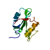
| ||||||||
|---|---|---|---|---|---|---|---|---|---|
| 1 |
| ||||||||
| Unit cell |
|
-Components
| #1: Protein | Mass: 12162.913 Da / Num. of mol.: 1 / Fragment: SH2 Domain (UNP residues 73 to 178) Source method: isolated from a genetically manipulated source Source: (gene. exp.) Homo sapiens (human) / Strain: BL21(DE3) / Gene: CSK / Plasmid: pTWIN1 / Production host:  References: UniProt: P41240, non-specific protein-tyrosine kinase |
|---|---|
| #2: Water | ChemComp-HOH /  Mass: 18.015 Da / Num. of mol.: 106 / Source method: isolated from a natural source / Formula: H2O Mass: 18.015 Da / Num. of mol.: 106 / Source method: isolated from a natural source / Formula: H2O |
| Has protein modification | Y |
-Experimental details
-Experiment
| Experiment | Method: X-RAY DIFFRACTION / Number of used crystals: 1 |
|---|
- Sample preparation
| Crystal | Density Matthews: 1.83 Å3/Da / Density % sol: 32.67 % |
|---|---|
| Crystal grow | Temperature: 298 K / Method: evaporation / pH: 7.3 Details: 22% PEG 4000, 100 mM Bis-Tris, pH 7.3., EVAPORATION, temperature 298K |
-Data collection
| Diffraction | Mean temperature: 80 K |
|---|---|
| Diffraction source | Source: SYNCHROTRON / Site: NSLS  / Beamline: X4C / Beamline: X4C |
| Detector | Type: MAR scanner 345 mm plate / Detector: IMAGE PLATE / Date: Mar 26, 2008 / Details: mirrors |
| Radiation | Protocol: SINGLE WAVELENGTH / Monochromatic (M) / Laue (L): M / Scattering type: x-ray |
| Radiation wavelength | Relative weight: 1 |
| Reflection | Resolution: 1.37→50 Å / Num. obs: 22360 / % possible obs: 99.8 % / Observed criterion σ(F): 2 / Observed criterion σ(I): 2 / Redundancy: 13.9 % / Biso Wilson estimate: 10.284 Å2 |
| Reflection shell | Highest resolution: 1.37 Å |
- Processing
| Software |
| |||||||||||||||||||||||||
|---|---|---|---|---|---|---|---|---|---|---|---|---|---|---|---|---|---|---|---|---|---|---|---|---|---|---|
| Refinement | Method to determine structure: MOLECULAR REPLACEMENT Starting model: PDB ENTRY 1K9A, sh2 part Resolution: 1.37→29.36 Å / Cor.coef. Fo:Fc: 0.95 / Cor.coef. Fo:Fc free: 0.952 / SU B: 1.053 / SU ML: 0.044 / Cross valid method: THROUGHOUT / ESU R: 0.067 / ESU R Free: 0.069 Stereochemistry target values: MAXIMUM LIKELIHOOD WITH PHASES Details: HYDROGENS HAVE BEEN ADDED IN THE RIDING POSITIONS
| |||||||||||||||||||||||||
| Solvent computation | Ion probe radii: 0.8 Å / Shrinkage radii: 0.8 Å / VDW probe radii: 1.2 Å / Solvent model: MASK | |||||||||||||||||||||||||
| Displacement parameters | Biso mean: 11.359 Å2
| |||||||||||||||||||||||||
| Refinement step | Cycle: LAST / Resolution: 1.37→29.36 Å
| |||||||||||||||||||||||||
| LS refinement shell | Resolution: 1.37→1.405 Å / Total num. of bins used: 20
|
