 Movie
Movie Controller

[English] 日本語
 Yorodumi
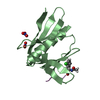
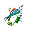
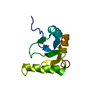
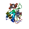
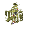
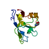
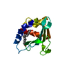
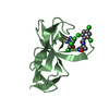
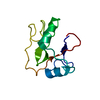
- PDB-2xi1: Crystal structure of the HIV-1 Nef sequenced from a patient's sample -
+ Open data

- Basic information
| Entry | Database: PDB / ID: 2xi1 | ||||||
|---|---|---|---|---|---|---|---|
| Title | Crystal structure of the HIV-1 Nef sequenced from a patient's sample | ||||||
 Components Components | (NEF) x 2 | ||||||
 Keywords Keywords | VIRAL PROTEIN / AIDS | ||||||
| Function / homology |  Function and homology information Function and homology informationsymbiont-mediated suppression of host antigen processing and presentation of peptide antigen via MHC class I / symbiont-mediated suppression of host antigen processing and presentation of peptide antigen via MHC class II / symbiont-mediated suppression of host autophagy / host cell Golgi membrane / SH3 domain binding / virion component / GTP binding / host cell plasma membrane / extracellular region / membrane Similarity search - Function | ||||||
| Biological species |   HUMAN IMMUNODEFICIENCY VIRUS TYPE 1 HUMAN IMMUNODEFICIENCY VIRUS TYPE 1 | ||||||
| Method |  X-RAY DIFFRACTION / OTHER / Resolution: 3.5 Å X-RAY DIFFRACTION / OTHER / Resolution: 3.5 Å | ||||||
 Authors Authors | Yadav, G.P. / Singh, P. / Gupta, S. / Tripathi, A.K. / Tripathi, R.K. / Ramachandran, R. | ||||||
 Citation Citation | Journal: Plos One / Year: 2011 Title: A Novel Dimer-Tetramer Transition Captured by the Crystal Structure of the HIV-1 Nef. Authors: Singh, P. / Yadav, G.P. / Gupta, S. / Tripathi, A.K. / Ramachandran, R. / Tripathi, R.K. | ||||||
| History |
|
- Structure visualization
| Structure viewer | Molecule: MolmilJmol/JSmol |
|---|
- Downloads & links
-Download
| PDBx/mmCIF format | 2xi1.cif.gz | 97.5 KB | Display | PDBx/mmCIF format |
|---|---|---|---|---|
| PDB format | pdb2xi1.ent.gz | 75.1 KB | Display | PDB format |
| PDBx/mmJSON format | 2xi1.json.gz | Tree view | PDBx/mmJSON format | |
| Others |  Other downloads Other downloads |
-Validation report
| Arichive directory | https://data.pdbj.org/pub/pdb/validation_reports/xi/2xi1ftp://data.pdbj.org/pub/pdb/validation_reports/xi/2xi1 | HTTPS FTP |
|---|
-Related structure data
| Similar structure data |
|---|

-Links
 PDBj
PDBj- Assembly
| Deposited unit | 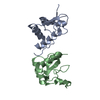
| ||||||||
|---|---|---|---|---|---|---|---|---|---|
| 1 | 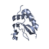
| ||||||||
| 2 | 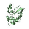
| ||||||||
| Unit cell |
|
-Components
| #1: Protein | Mass: 23612.531 Da / Num. of mol.: 1 Source method: isolated from a genetically manipulated source Source: (gene. exp.) HUMAN IMMUNODEFICIENCY VIRUS TYPE 1 (BH5 ISOLATE)Description: PATIENT SAMPLE / Production host:  |
|---|---|
| #2: Protein | Mass: 23612.598 Da / Num. of mol.: 1 Source method: isolated from a genetically manipulated source Source: (gene. exp.) HUMAN IMMUNODEFICIENCY VIRUS TYPE 1 (BH5 ISOLATE)Description: PATIENT SAMPLE / Production host: |
| Sequence details | GENBANK ACCESSION NUMBER GQ184340 CORRESPOND |
-Experimental details
-Experiment
| Experiment | Method: X-RAY DIFFRACTION / Number of used crystals: 1 |
|---|
- Sample preparation
| Crystal | Density Matthews: 3.02 Å3/Da / Density % sol: 59.22 % / Description: NONE |
|---|
-Data collection
| Diffraction | Mean temperature: 100 K |
|---|---|
| Diffraction source | Source: ROTATING ANODE / Type: RIGAKU MICROMAX-007 HF / Wavelength: 1.5418 |
| Detector | Type: MAR scanner 345 mm plate / Detector: IMAGE PLATE / Details: VARIMAX-HF |
| Radiation | Protocol: SINGLE WAVELENGTH / Monochromatic (M) / Laue (L): M / Scattering type: x-ray |
| Radiation wavelength | Wavelength: 1.5418 Å / Relative weight: 1 |
| Reflection | Resolution: 3.5→29.9 Å / Num. obs: 7262 / % possible obs: 93 % / Observed criterion σ(I): 2 / Redundancy: 5.7 % / Biso Wilson estimate: 56.37 Å2 / Rmerge(I) obs: 0.12 / Net I/σ(I): 7.1 |
- Processing
| Software |
| ||||||||||||||||||||||||||||||||||||||||
|---|---|---|---|---|---|---|---|---|---|---|---|---|---|---|---|---|---|---|---|---|---|---|---|---|---|---|---|---|---|---|---|---|---|---|---|---|---|---|---|---|---|
| Refinement | Method to determine structure: OTHER Starting model: NONE Resolution: 3.5→29.551 Å / SU ML: 0.45 / σ(F): 1.36 / Phase error: 26.11 / Stereochemistry target values: ML / Details: 1-75 AND 152-178 ARE DISORDERED
| ||||||||||||||||||||||||||||||||||||||||
| Solvent computation | Shrinkage radii: 0.9 Å / VDW probe radii: 1.11 Å / Solvent model: FLAT BULK SOLVENT MODEL / Bsol: 34.873 Å2 / ksol: 0.335 e/Å3 | ||||||||||||||||||||||||||||||||||||||||
| Displacement parameters | Biso mean: 37 Å2
| ||||||||||||||||||||||||||||||||||||||||
| Refinement step | Cycle: LAST / Resolution: 3.5→29.551 Å
| ||||||||||||||||||||||||||||||||||||||||
| Refine LS restraints |
| ||||||||||||||||||||||||||||||||||||||||
| LS refinement shell |
| ||||||||||||||||||||||||||||||||||||||||
| Refinement TLS params. | Method: refined / Origin x: -35.9128 Å / Origin y: 40.169 Å / Origin z: 10.8204 Å
| ||||||||||||||||||||||||||||||||||||||||
| Refinement TLS group | Selection details: ALL |
