 Movie
Movie Controller

[English] 日本語
 Yorodumi
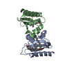
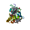
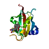
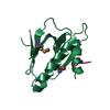
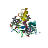
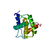
- PDB-3bks: Crystal Structure of Sterol Carrier Protein-2 like-3 (SCP2-L3) fr... -
+ Open data

- Basic information
| Entry | Database: PDB / ID: 3bks | ||||||
|---|---|---|---|---|---|---|---|
| Title | Crystal Structure of Sterol Carrier Protein-2 like-3 (SCP2-L3) from Aedes Aegypti | ||||||
 Components Components | Sterol Carrier Protein-2 like-3 | ||||||
 Keywords Keywords | LIPID BINDING PROTEIN / Sterol Carrier / Mosquito / Fatty Acid / Palmitic acid / cholesterol | ||||||
| Function / homology | SCP2 sterol-binding domain / SCP-2 sterol transfer family / SCP2 sterol-binding domain / Nonspecific Lipid-transfer Protein; Chain A / SCP2 sterol-binding domain superfamily / 2-Layer Sandwich / Alpha Beta / PALMITIC ACID / AAEL012704-PA Function and homology information Function and homology information | ||||||
| Biological species |  | ||||||
| Method |  X-RAY DIFFRACTION / MOLECULAR REPLACEMENT / Resolution: 2.1 Å X-RAY DIFFRACTION / MOLECULAR REPLACEMENT / Resolution: 2.1 Å | ||||||
 Authors Authors | Dyer, D.H. / Lan, Q. / Forest, K.T. | ||||||
 Citation Citation | Journal: Mol.Cell.Biochem. / Year: 2009 Title: Characterization of the yellow fever mosquito sterol carrier protein-2 like 3 gene and ligand-bound protein structure. Authors: Dyer, D.H. / Vyazunova, I. / Lorch, J.M. / Forest, K.T. / Lan, Q. | ||||||
| History |
|
- Structure visualization
| Structure viewer | Molecule: MolmilJmol/JSmol |
|---|
- Downloads & links
-Download
| PDBx/mmCIF format | 3bks.cif.gz | 38.1 KB | Display | PDBx/mmCIF format |
|---|---|---|---|---|
| PDB format | pdb3bks.ent.gz | 25.5 KB | Display | PDB format |
| PDBx/mmJSON format | 3bks.json.gz | Tree view | PDBx/mmJSON format | |
| Others |  Other downloads Other downloads |
-Validation report
| Arichive directory | https://data.pdbj.org/pub/pdb/validation_reports/bk/3bksftp://data.pdbj.org/pub/pdb/validation_reports/bk/3bks | HTTPS FTP |
|---|
-Related structure data
| Related structure data |  3bkrC  1pz4S S: Starting model for refinement C: citing same article ( |
|---|---|
| Similar structure data |


-Links
 PDBj
PDBj


- Assembly
| Deposited unit | 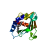
| ||||||||
|---|---|---|---|---|---|---|---|---|---|
| 1 |
| ||||||||
| Unit cell |
|
-Components
| #1: Protein | Mass: 14181.137 Da / Num. of mol.: 1 Source method: isolated from a genetically manipulated source Source: (gene. exp.)  |
|---|---|
| #2: Chemical | ChemComp-PLM /   Mass: 256.424 Da / Num. of mol.: 1 / Source method: obtained synthetically / Formula: C16H32O2 Mass: 256.424 Da / Num. of mol.: 1 / Source method: obtained synthetically / Formula: C16H32O2 |
| #3: Water | ChemComp-HOH /  Mass: 18.015 Da / Num. of mol.: 41 / Source method: isolated from a natural source / Formula: H2O Mass: 18.015 Da / Num. of mol.: 41 / Source method: isolated from a natural source / Formula: H2O |
-Experimental details
-Experiment
| Experiment | Method: X-RAY DIFFRACTION / Number of used crystals: 1 |
|---|
- Sample preparation
| Crystal | Density Matthews: 2.79 Å3/Da / Density % sol: 55.91 % |
|---|---|
| Crystal grow | Temperature: 296 K / Method: vapor diffusion, hanging drop / pH: 7.5 Details: 2 microl protein at 10mg/ml was mixed with 2microl mother liquor consisting of 25% Peg 8000, 200mM AmSO4, 200mM AmF, pH 7.5, VAPOR DIFFUSION, HANGING DROP, temperature 296K |
-Data collection
| Diffraction | Mean temperature: 296 K |
|---|---|
| Diffraction source | Source: ROTATING ANODE / Type: BRUKER AXS MICROSTAR / Wavelength: 1.54 Å |
| Detector | Type: Bruker Proteum / Date: Jan 22, 2007 |
| Radiation | Monochromator: graded mirrors / Protocol: SINGLE WAVELENGTH / Monochromatic (M) / Laue (L): M / Scattering type: x-ray |
| Radiation wavelength | Wavelength: 1.54 Å / Relative weight: 1 |
| Reflection | Resolution: 2.1→25 Å / Num. all: 17937 / Num. obs: 9644 / % possible obs: 99.9 % / Redundancy: 1.9 % / Biso Wilson estimate: 22.5 Å2 / Rsym value: 0.018 / Net I/σ(I): 38.3 |
| Reflection shell | Resolution: 2.1→2.2 Å / Redundancy: 1.9 % / Mean I/σ(I) obs: 5.9 / Num. unique all: 1232 / Rsym value: 0.063 / % possible all: 99 |
- Processing
| Software |
| ||||||||||||||||||||||||||||||||||||||||||||||||||||||||||||||||||||||||||||||||||||||||||
|---|---|---|---|---|---|---|---|---|---|---|---|---|---|---|---|---|---|---|---|---|---|---|---|---|---|---|---|---|---|---|---|---|---|---|---|---|---|---|---|---|---|---|---|---|---|---|---|---|---|---|---|---|---|---|---|---|---|---|---|---|---|---|---|---|---|---|---|---|---|---|---|---|---|---|---|---|---|---|---|---|---|---|---|---|---|---|---|---|---|---|---|
| Refinement | Method to determine structure: MOLECULAR REPLACEMENT Starting model: Polyalanine model derived from 1PZ4 Resolution: 2.1→25 Å / Cor.coef. Fo:Fc: 0.96 / Cor.coef. Fo:Fc free: 0.948 / SU B: 6.526 / SU ML: 0.087 / Cross valid method: THROUGHOUT / σ(F): 0 / ESU R: 0.159 / ESU R Free: 0.135 / Stereochemistry target values: MAXIMUM LIKELIHOOD
| ||||||||||||||||||||||||||||||||||||||||||||||||||||||||||||||||||||||||||||||||||||||||||
| Solvent computation | Ion probe radii: 0.8 Å / Shrinkage radii: 0.8 Å / VDW probe radii: 1.4 Å / Solvent model: MASK | ||||||||||||||||||||||||||||||||||||||||||||||||||||||||||||||||||||||||||||||||||||||||||
| Displacement parameters | Biso mean: 14.127 Å2
| ||||||||||||||||||||||||||||||||||||||||||||||||||||||||||||||||||||||||||||||||||||||||||
| Refine analyze |
| ||||||||||||||||||||||||||||||||||||||||||||||||||||||||||||||||||||||||||||||||||||||||||
| Refinement step | Cycle: LAST / Resolution: 2.1→25 Å
| ||||||||||||||||||||||||||||||||||||||||||||||||||||||||||||||||||||||||||||||||||||||||||
| Refine LS restraints |
| ||||||||||||||||||||||||||||||||||||||||||||||||||||||||||||||||||||||||||||||||||||||||||
| LS refinement shell | Resolution: 2.1→2.2 Å / Total num. of bins used: 20
|
