[English] 日本語
Yorodumi
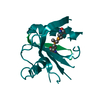
- PDB-2g43: Structure of the ZNF UBP domain from deubiquitinating enzyme isop... -

+
Open data


ID or keywords:

Loading...

-
Basic information

Entry
Database: PDB / ID: 2g43
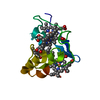
TitleStructure of the ZNF UBP domain from deubiquitinating enzyme isopeptidase T (IsoT)
ComponentsUbiquitin carboxyl-terminal hydrolase 5
KeywordsHYDROLASE / Zinc Finger / deubiquitinating enzyme
Function / homology
Function and homology information


protein K48-linked deubiquitination / deubiquitinase activity / negative regulation of ubiquitin-dependent protein catabolic process / Synthesis of active ubiquitin: roles of E1 and E2 enzymes / ubiquitin binding / positive regulation of proteasomal ubiquitin-dependent protein catabolic process / ubiquitinyl hydrolase 1 / cysteine-type deubiquitinase activity / lysosome / Ub-specific processing proteases ...protein K48-linked deubiquitination / deubiquitinase activity / negative regulation of ubiquitin-dependent protein catabolic process / Synthesis of active ubiquitin: roles of E1 and E2 enzymes / ubiquitin binding / positive regulation of proteasomal ubiquitin-dependent protein catabolic process / ubiquitinyl hydrolase 1 / cysteine-type deubiquitinase activity / lysosome / Ub-specific processing proteases / protein ubiquitination / cysteine-type endopeptidase activity / proteolysis / zinc ion binding / nucleus / cytosol
Similarity search - Function
Ubiquitin carboxyl-terminal hydrolase 5, UBA1 domain / Ubiquitinyl hydrolase / Ubiquitinyl hydrolase, variant UBP zinc finger / Variant UBP zinc finger / : / Ubiquitin Carboxyl-terminal Hydrolase-like zinc finger / Zinc finger, UBP-type / Zn-finger in ubiquitin-hydrolases and other protein / Zinc finger UBP-type profile. / UBA/TS-N domain ...Ubiquitin carboxyl-terminal hydrolase 5, UBA1 domain / Ubiquitinyl hydrolase / Ubiquitinyl hydrolase, variant UBP zinc finger / Variant UBP zinc finger / : / Ubiquitin Carboxyl-terminal Hydrolase-like zinc finger / Zinc finger, UBP-type / Zn-finger in ubiquitin-hydrolases and other protein / Zinc finger UBP-type profile. / UBA/TS-N domain / Ubiquitin specific protease (USP) domain signature 2. / Ubiquitin specific protease (USP) domain signature 1. / Ubiquitin specific protease, conserved site / Ubiquitin associated domain / Peptidase C19, ubiquitin carboxyl-terminal hydrolase / Ubiquitin carboxyl-terminal hydrolase / Ubiquitin specific protease domain / Ubiquitin specific protease (USP) domain profile. / Ubiquitin-associated domain / Ubiquitin-associated domain (UBA) profile. / UBA-like superfamily / Zinc/RING finger domain, C3HC4 (zinc finger) / Herpes Virus-1 / Papain-like cysteine peptidase superfamily / Zinc finger, RING/FYVE/PHD-type / 2-Layer Sandwich / Alpha Beta
Similarity search - Domain/homology
Ubiquitin carboxyl-terminal hydrolase 5
Similarity search - Component
Biological speciesHomo sapiens (human)
MethodX-RAY DIFFRACTION / SYNCHROTRON / MAD / Resolution: 2.09 Å
AuthorsReyes-Turcu, F.E. / Horton, J.R. / Mullally, J.E. / Heroux, A. / Cheng, X. / Wilkinson, K.D.
CitationJournal: Cell(Cambridge,Mass.) / Year: 2006
Title: The Ubiquitin Binding Domain ZnF UBP Recognizes the C-Terminal Diglycine Motif of Unanchored Ubiquitin.
Authors: Reyes-Turcu, F.E. / Horton, J.R. / Mullally, J.E. / Heroux, A. / Cheng, X. / Wilkinson, K.D.
History
DepositionFeb 21, 2006Deposition site: RCSB / Processing site: RCSB
Revision 1.0Apr 4, 2006Provider: repository / Type: Initial release
Revision 1.1May 1, 2008Group: Version format compliance
Revision 1.2Jul 13, 2011Group: Version format compliance
Remark 600HETEROGEN THREE UNKOWN ATOMS, UNX 61, 65 AND 66 FORM A TRIANGULAR STRUCTURE.

-
Structure visualization

Structure viewerMolecule:
MolmilJmol/JSmol

Downloads & links

-
Assembly

Deposited unit
A: Ubiquitin carboxyl-terminal hydrolase 5
B: Ubiquitin carboxyl-terminal hydrolase 5
hetero molecules


Theoretical massNumber of molelcules
Total (without water)29,3187
Polymers29,1872
Non-polymers1315
Water1,27971
1
A: Ubiquitin carboxyl-terminal hydrolase 5
hetero molecules


Theoretical massNumber of molelcules
Total (without water)14,6592
Polymers14,5931
Non-polymers651
Water181
TypeNameSymmetry operationNumber
identity operation1_555x,y,z1
2
B: Ubiquitin carboxyl-terminal hydrolase 5
hetero molecules


Theoretical massNumber of molelcules
Total (without water)14,6595
Polymers14,5931
Non-polymers654
Water181
TypeNameSymmetry operationNumber
identity operation1_555x,y,z1
Unit cell
Length a, b, c (Å)61.380, 86.180, 59.900
Angle α, β, γ (deg.)90.00, 99.29, 90.00
Int Tables number5
Space group name H-MC121

-
Components

#1: Protein Ubiquitin carboxyl-terminal hydrolase 5 / Ubiquitin thiolesterase 5 / Ubiquitin-specific processing protease 5 / Deubiquitinating enzyme 5 / ...Ubiquitin thiolesterase 5 / Ubiquitin-specific processing protease 5 / Deubiquitinating enzyme 5 / Isopeptidase T


Mass: 14593.387 Da / Num. of mol.: 2
Fragment: The Zn Finger UMP domain of IsoT (residues 163-291)
Source method: isolated from a genetically manipulated source
Source: (gene. exp.) Homo sapiens (human) / Gene: USP5, ISOT / Production host: Escherichia coli (E. coli) / References: UniProt: P45974, EC: 3.1.2.15
#2: Chemical ChemComp-ZN / ZINC ION


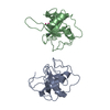
Mass: 65.409 Da / Num. of mol.: 2 / Source method: obtained synthetically / Formula: Zn
#3: Chemical ChemComp-UNX / UNKNOWN ATOM OR ION


Num. of mol.: 3 / Source method: obtained synthetically
#4: Water ChemComp-HOH / water


Mass: 18.015 Da / Num. of mol.: 71 / Source method: isolated from a natural source / Formula: H2O

-
Experimental details

-
Experiment

ExperimentMethod: X-RAY DIFFRACTION / Number of used crystals: 2

-
Sample preparation

CrystalDensity Matthews: 2.68 Å3/Da / Density % sol: 54.06 %
Crystal growTemperature: 300 K / pH: 6.5
Details: 16% polyethylene glycol 8000, 0.8 M sodium cacodylate pH 6.5, 0.16 M calcium acetate, 20% glycerol, VAPOR DIFFUSION, HANGING DROP, temperature 300K, pH 6.50

-
Data collection

Diffraction
IDMean temperature (K)Crystal-ID
11001
21
Diffraction sourceSource: SYNCHROTRON / Site: NSLS / Beamline: X26C / Wavelength: 1.28020, 1.2790, 1.12810 0.97623
DetectorType: ADSC QUANTUM 4 / Detector: CCD / Date: May 5, 2005
Radiation
IDMonochromatorProtocolMonochromatic (M) / Laue (L)Scattering typeWavelength-ID
1NSLS X26CMADMx-ray1
2Mx-ray1
Radiation wavelength
IDWavelength (Å)Relative weight
11.28021
21.2791
31.12811
40.976231
ReflectionResolution: 2.09→35 Å / Num. obs: 17788 / % possible obs: 96.7 % / Observed criterion σ(I): -3 / Redundancy: 2.8 % / Biso Wilson estimate: 21.2 Å2 / Rmerge(I) obs: 0.047 / Net I/σ(I): 27.7
Reflection shellResolution: 2.09→2.16 Å / Rmerge(I) obs: 0.064 / Mean I/σ(I) obs: 16.7 / % possible all: 97.2

-
Processing

Software
NameVersionClassification
HKL-2000data collection
SCALEPACKdata scaling
SOLVEphasing
CNS1.1refinement
HKL-2000data reduction
RefinementMethod to determine structure: MAD / Resolution: 2.09→35 Å / Rfactor Rfree error: 0.006 / Data cutoff high absF: 445910.92 / Data cutoff low absF: 0 / Isotropic thermal model: OVERALL ANISOTROPIC B VALUE / Cross valid method: THROUGHOUT / σ(F): 0 / Stereochemistry target values: ENGH & HUBER
RfactorNum. reflection% reflectionSelection details
Rfree0.274 1743 5 %RANDOM
Rwork0.228 ---
obs0.228 17788 96.7 %-
all-17788 --
Solvent computationSolvent model: FLAT MODEL / Bsol: 46.06 Å2 / ksol: 0.39 e/Å3
Displacement parametersBiso mean: 41.5 Å2
Baniso -1Baniso -2Baniso -3
1--7.05 Å20 Å21.41 Å2
2--3.72 Å20 Å2
3---3.33 Å2
Refine analyze
FreeObs
Luzzati coordinate error0.33 Å0.3 Å
Luzzati d res low-35 Å
Luzzati sigma a0.2 Å0.21 Å
Refinement stepCycle: LAST / Resolution: 2.09→35 Å
ProteinNucleic acidLigandSolventTotal
Num. atoms1757 0 5 71 1833
Refine LS restraints
Refine-IDTypeDev idealDev ideal target
X-RAY DIFFRACTIONc_bond_d0.005
X-RAY DIFFRACTIONc_bond_d_na
X-RAY DIFFRACTIONc_bond_d_prot
X-RAY DIFFRACTIONc_angle_d
X-RAY DIFFRACTIONc_angle_d_na
X-RAY DIFFRACTIONc_angle_d_prot
X-RAY DIFFRACTIONc_angle_deg1.4
X-RAY DIFFRACTIONc_angle_deg_na
X-RAY DIFFRACTIONc_angle_deg_prot
X-RAY DIFFRACTIONc_dihedral_angle_d24.2
X-RAY DIFFRACTIONc_dihedral_angle_d_na
X-RAY DIFFRACTIONc_dihedral_angle_d_prot
X-RAY DIFFRACTIONc_improper_angle_d0.82
X-RAY DIFFRACTIONc_improper_angle_d_na
X-RAY DIFFRACTIONc_improper_angle_d_prot
X-RAY DIFFRACTIONc_mcbond_it1.581.5
X-RAY DIFFRACTIONc_mcangle_it2.642
X-RAY DIFFRACTIONc_scbond_it2.452
X-RAY DIFFRACTIONc_scangle_it3.792.5
LS refinement shellResolution: 2.09→2.16 Å / Rfactor Rfree error: 0.025 / Total num. of bins used: 10
RfactorNum. reflection% reflection
Rfree0.3 146 4.5 %
Rwork0.289 3097 -
obs--89.3 %
Xplor file
Refine-IDSerial noParam fileTopol file
X-RAY DIFFRACTION1protein_rep.paramprotein.top
X-RAY DIFFRACTION2dna-rna_rep.param
X-RAY DIFFRACTION4water_rep.param
X-RAY DIFFRACTION5ion.param

+
About Yorodumi

-
News

-
Feb 9, 2022. New format data for meta-information of EMDB entries

New format data for meta-information of EMDB entries

  • Version 3 of the EMDB header file is now the official format.
  • The previous official version 1.9 will be removed from the archive.

Related info.:EMDB header

External links:wwPDB to switch to version 3 of the EMDB data model

-
Aug 12, 2020. Covid-19 info

Covid-19 info

URL: https://pdbjlc1.pdbj.org/emnavi/covid19.php

New page: Covid-19 featured information page in EM Navigator.

Related info.:Covid-19 info / Mar 5, 2020. Novel coronavirus structure data

+
Mar 5, 2020. Novel coronavirus structure data

Novel coronavirus structure data

Related info.:Yorodumi Speices / Aug 12, 2020. Covid-19 info

External links:COVID-19 featured content - PDBj / Molecule of the Month (242):Coronavirus Proteases

+
Jan 31, 2019. EMDB accession codes are about to change! (news from PDBe EMDB page)

EMDB accession codes are about to change! (news from PDBe EMDB page)

  • The allocation of 4 digits for EMDB accession codes will soon come to an end. Whilst these codes will remain in use, new EMDB accession codes will include an additional digit and will expand incrementally as the available range of codes is exhausted. The current 4-digit format prefixed with “EMD-” (i.e. EMD-XXXX) will advance to a 5-digit format (i.e. EMD-XXXXX), and so on. It is currently estimated that the 4-digit codes will be depleted around Spring 2019, at which point the 5-digit format will come into force.
  • The EM Navigator/Yorodumi systems omit the EMD- prefix.

Related info.:Q: What is EMD? / ID/Accession-code notation in Yorodumi/EM Navigator

External links:EMDB Accession Codes are Changing Soon! / Contact to PDBj

+
Jul 12, 2017. Major update of PDB

Major update of PDB

  • wwPDB released updated PDB data conforming to the new PDBx/mmCIF dictionary.
  • This is a major update changing the version number from 4 to 5, and with Remediation, in which all the entries are updated.
  • In this update, many items about electron microscopy experimental information are reorganized (e.g. em_software).
  • Now, EM Navigator and Yorodumi are based on the updated data.

External links:wwPDB Remediation / Enriched Model Files Conforming to OneDep Data Standards Now Available in the PDB FTP Archive

-
Yorodumi

Thousand views of thousand structures

  • Yorodumi is a browser for structure data from EMDB, PDB, SASBDB, etc.
  • This page is also the successor to EM Navigator detail page, and also detail information page/front-end page for Omokage search.
  • The word "yorodu" (or yorozu) is an old Japanese word meaning "ten thousand". "mi" (miru) is to see.

Related info.:EMDB / PDB / SASBDB / Comparison of 3 databanks / Yorodumi Search / Aug 31, 2016. New EM Navigator & Yorodumi / Yorodumi Papers / Jmol/JSmol / Function and homology information / Changes in new EM Navigator and Yorodumi

Read more