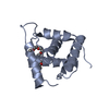
 Movie
Movie Controller


+ Open data

- Basic information
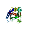
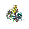
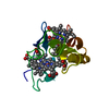
| Entry | Database: PDB / ID: 1i77 | ||||||
|---|---|---|---|---|---|---|---|
| Title | CYTOCHROME C3 FROM DESULFOVIBRIO DESULFURICANS ESSEX 6 | ||||||
 Components Components | CYTOCHROME C3 | ||||||
 Keywords Keywords | ELECTRON TRANSPORT / multi-heme cytochrome c | ||||||
| Function / homology |  Function and homology information Function and homology information | ||||||
| Biological species |  Desulfovibrio desulfuricans (bacteria) Desulfovibrio desulfuricans (bacteria) | ||||||
| Method |  X-RAY DIFFRACTION / MOLECULAR REPLACEMENT / Resolution: 1.95 Å X-RAY DIFFRACTION / MOLECULAR REPLACEMENT / Resolution: 1.95 Å | ||||||
 Authors Authors | Einsle, O. / Foerster, S. / Mann, K.H. / Fritz, G. / Messerschmidt, A. / Kroneck, P.M.H. | ||||||
 Citation Citation | Journal: Eur.J.Biochem. / Year: 2001 Title: Spectroscopic investigation and determination of reactivity and structure of the tetraheme cytochrome c3 from Desulfovibrio desulfuricans Essex 6. Authors: Einsle, O. / Foerster, S. / Mann, K. / Fritz, G. / Messerschmidt, A. / Kroneck, P.M. | ||||||
| History |
|
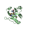
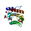
- Structure visualization
| Structure viewer | Molecule: MolmilJmol/JSmol |
|---|
- Downloads & links
-Download
| PDBx/mmCIF format | 1i77.cif.gz | 40.7 KB | Display | PDBx/mmCIF format |
|---|---|---|---|---|
| PDB format | pdb1i77.ent.gz | 28.3 KB | Display | PDB format |
| PDBx/mmJSON format | 1i77.json.gz | Tree view | PDBx/mmJSON format | |
| Others |  Other downloads Other downloads |
-Validation report
| Arichive directory | https://data.pdbj.org/pub/pdb/validation_reports/i7/1i77ftp://data.pdbj.org/pub/pdb/validation_reports/i7/1i77 | HTTPS FTP |
|---|
-Related structure data
| Related structure data |  2cyr S: Starting model for refinement |
|---|---|
| Similar structure data |


-Links
 PDBj
PDBj
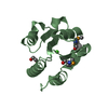
- Assembly
| Deposited unit | 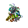
| ||||||||
|---|---|---|---|---|---|---|---|---|---|
| 1 |
| ||||||||
| Unit cell |
|
-Components
| #1: Protein | Mass: 11637.477 Da / Num. of mol.: 1 / Source method: isolated from a natural source / Source: (natural) Desulfovibrio desulfuricans (bacteria) / Strain: ESSEX 6 / References: UniProt: Q9L915 | ||||
|---|---|---|---|---|---|
| #2: Chemical | ChemComp-HEM /   Mass: 616.487 Da / Num. of mol.: 4 / Source method: obtained synthetically / Formula: C34H32FeN4O4 Mass: 616.487 Da / Num. of mol.: 4 / Source method: obtained synthetically / Formula: C34H32FeN4O4#3: Water | ChemComp-HOH / |  Mass: 18.015 Da / Num. of mol.: 117 / Source method: isolated from a natural source / Formula: H2O Mass: 18.015 Da / Num. of mol.: 117 / Source method: isolated from a natural source / Formula: H2OHas protein modification | Y | |
-Experimental details
-Experiment
| Experiment | Method: X-RAY DIFFRACTION / Number of used crystals: 1 |
|---|
- Sample preparation
| Crystal | Density Matthews: 2.74 Å3/Da / Density % sol: 55.08 % | |||||||||||||||||||||||||
|---|---|---|---|---|---|---|---|---|---|---|---|---|---|---|---|---|---|---|---|---|---|---|---|---|---|---|
| Crystal grow | Temperature: 293 K / Method: vapor diffusion, sitting drop / pH: 4.6 Details: PEG MME 2000, sodium acetate, ammonium sulfate, pH 4.6, VAPOR DIFFUSION, SITTING DROP, temperature 293K | |||||||||||||||||||||||||
| Crystal grow | *PLUS Temperature: 21 ℃ | |||||||||||||||||||||||||
| Components of the solutions | *PLUS
|
-Data collection
| Diffraction | Mean temperature: 293 K |
|---|---|
| Diffraction source | Source: ROTATING ANODE / Type: RIGAKU / Wavelength: 1.5418 Å |
| Detector | Type: MARRESEARCH / Detector: IMAGE PLATE / Date: Jul 3, 1997 |
| Radiation | Monochromator: GRAPHITE / Protocol: SINGLE WAVELENGTH / Monochromatic (M) / Laue (L): M / Scattering type: x-ray |
| Radiation wavelength | Wavelength: 1.5418 Å / Relative weight: 1 |
| Reflection | Resolution: 1.95→18 Å / Num. all: 9821 / Num. obs: 9654 / % possible obs: 98.3 % / Observed criterion σ(F): 3 / Observed criterion σ(I): 3 / Redundancy: 3.8 % / Biso Wilson estimate: 25.98 Å2 / Rmerge(I) obs: 0.117 / Net I/σ(I): 9.9 |
| Reflection shell | Resolution: 1.95→2.01 Å / Redundancy: 3.6 % / Rmerge(I) obs: 0.335 / Mean I/σ(I) obs: 3.1 / % possible all: 98.5 |
| Reflection | *PLUS Num. measured all: 59364 |
| Reflection shell | *PLUS % possible obs: 98.5 % |
- Processing
| Software |
| |||||||||||||||||||||||||
|---|---|---|---|---|---|---|---|---|---|---|---|---|---|---|---|---|---|---|---|---|---|---|---|---|---|---|
| Refinement | Method to determine structure: MOLECULAR REPLACEMENT Starting model: PDB ENTRY 2CYR 2cyr Resolution: 1.95→18 Å / Isotropic thermal model: Isotropic / σ(F): 2 / Stereochemistry target values: Engh & Huber
| |||||||||||||||||||||||||
| Displacement parameters | Biso mean: 29.92 Å2
| |||||||||||||||||||||||||
| Refinement step | Cycle: LAST / Resolution: 1.95→18 Å
| |||||||||||||||||||||||||
| Refine LS restraints |
| |||||||||||||||||||||||||
| Software | *PLUS Name: CNS / Version: 1 / Classification: refinement | |||||||||||||||||||||||||
| Refinement | *PLUS Lowest resolution: 18 Å / σ(F): 2 / Rfactor obs: 0.173 | |||||||||||||||||||||||||
| Solvent computation | *PLUS | |||||||||||||||||||||||||
| Displacement parameters | *PLUS |
