SEQUENCE THE CONSTRUCT WAS EXPRESSED WITH A PURIFICATION TAG MGSDKIHHHHHHENLYFQG. THE TAG WAS ... SEQUENCE THE CONSTRUCT WAS EXPRESSED WITH A PURIFICATION TAG MGSDKIHHHHHHENLYFQG. THE TAG WAS REMOVED WITH TEV PROTEASE LEAVING ONLY A GLYCINE FOLLOWED BY THE TARGET SEQUENCE.
Monochromator: Single crystal Si(111) bent (horizontal focusing) Protocol: MAD / Monochromatic (M) / Laue (L): M / Scattering type: x-ray
Radiation wavelength
ID
Wavelength (Å)
Relative weight
1
0.91162
1
2
0.97929
1
Reflection
Resolution: 1.44→39.193 Å / Num. obs: 40271 / % possible obs: 77.4 % / Biso Wilson estimate: 25.909 Å2 / Rmerge(I) obs: 0.036 / Net I/σ(I): 14.96
Reflection shell
Diffraction-ID: 1
Resolution (Å)
Highest resolution (Å)
Rmerge(I) obs
Mean I/σ(I) obs
Num. measured obs
Num. unique all
% possible all
1.44-1.49
0.467
2.2
3810
1355
27.2
1.49-1.55
0.362
3
5952
2030
39
1.55-1.62
0.27
3.9
8354
2847
55.7
1.62-1.71
0.201
5.2
13321
4498
82.9
1.71-1.81
0.149
6.9
13315
4448
93.2
1.81-1.95
0.088
10.8
14593
4891
94.2
1.95-2.15
0.055
16.1
14803
4966
95.2
2.15-2.46
0.039
21.9
14552
4892
95.2
2.46
0.032
26.2
14540
4918
95.9
-
Phasing
Phasing
Method: MAD
-
Processing
Software
Name
Version
Classification
NB
MolProbity
3beta29
modelbuilding
SHELX
phasing
REFMAC
5.2.0019
refinement
XSCALE
datascaling
PDB_EXTRACT
2
dataextraction
MAR345
CCD
datacollection
XDS
datareduction
SHELXD
phasing
autoSHARP
phasing
Refinement
Method to determine structure: MAD / Resolution: 1.44→39.193 Å / Cor.coef. Fo:Fc: 0.973 / Cor.coef. Fo:Fc free: 0.959 / SU B: 3.311 / SU ML: 0.059 / TLS residual ADP flag: LIKELY RESIDUAL / Cross valid method: THROUGHOUT / σ(F): 0 / ESU R: 0.077 / ESU R Free: 0.079 Stereochemistry target values: MAXIMUM LIKELIHOOD WITH PHASES Details: 1. HYDROGENS HAVE BEEN ADDED IN THE RIDING POSITIONS. 2. A MET-INHIBITION PROTOCOL WAS USED FOR SELENOMETHIONINE INCORPORATION DURING PROTEIN EXPRESSION. THE OCCUPANCY OF THE SE ATOMS IN THE ...Details: 1. HYDROGENS HAVE BEEN ADDED IN THE RIDING POSITIONS. 2. A MET-INHIBITION PROTOCOL WAS USED FOR SELENOMETHIONINE INCORPORATION DURING PROTEIN EXPRESSION. THE OCCUPANCY OF THE SE ATOMS IN THE MSE RESIDUES WAS REDUCED TO 0.75 FOR THE REDUCED SCATTERING POWER DUE TO PARTIAL S-MET INCORPORATION. 3. ATOM RECORD CONTAINS RESIDUAL B FACTORS ONLY. 4. CALCIUM IS MODELED BASED ON CRYSTALLIZATION CONDITIONS AND COORDINATION ENVIRONMENT AND GEOMETRY. THE OCCUPANCIES ARE SET AS 0.5 ACCORDING TO DENSITY. STRUCTURAL HOMOLOGS CONTAIN MAGNESIUM AT THE CORRESPONDING SITES. 5. EDO MOLECULES ARE FROM CRYOPROTECTANT. 6. THE NOMINAL RESOLUTION IS 1.60 A WITH 5615 OBSERVED REFLECTIONS BETWEEN 1.60-1.44 (40.5% COMPLETE FOR THIS SHELL) INCLUDED IN THE REFINEMENT.
Rfactor
Num. reflection
% reflection
Selection details
Rfree
0.199
2034
5.1 %
RANDOM
Rwork
0.168
-
-
-
all
0.17
-
-
-
obs
0.17
40270
78.61 %
-
Solvent computation
Ion probe radii: 0.8 Å / Shrinkage radii: 0.8 Å / VDW probe radii: 1.2 Å / Solvent model: BABINET MODEL WITH MASK
In the structure databanks used in Yorodumi, some data are registered as the other names, "COVID-19 virus" and "2019-nCoV". Here are the details of the virus and the list of structure data.
Jan 31, 2019. EMDB accession codes are about to change! (news from PDBe EMDB page)
EMDB accession codes are about to change! (news from PDBe EMDB page)
The allocation of 4 digits for EMDB accession codes will soon come to an end. Whilst these codes will remain in use, new EMDB accession codes will include an additional digit and will expand incrementally as the available range of codes is exhausted. The current 4-digit format prefixed with “EMD-” (i.e. EMD-XXXX) will advance to a 5-digit format (i.e. EMD-XXXXX), and so on. It is currently estimated that the 4-digit codes will be depleted around Spring 2019, at which point the 5-digit format will come into force.
The EM Navigator/Yorodumi systems omit the EMD- prefix.
Related info.:Q: What is EMD? / ID/Accession-code notation in Yorodumi/EM Navigator
Yorodumi is a browser for structure data from EMDB, PDB, SASBDB, etc.
This page is also the successor to EM Navigator detail page, and also detail information page/front-end page for Omokage search.
The word "yorodu" (or yorozu) is an old Japanese word meaning "ten thousand". "mi" (miru) is to see.
Related info.:EMDB / PDB / SASBDB / Comparison of 3 databanks / Yorodumi Search / Aug 31, 2016. New EM Navigator & Yorodumi / Yorodumi Papers / Jmol/JSmol / Function and homology information / Changes in new EM Navigator and Yorodumi
 Movie
Movie Controller

 Yorodumi
 Open data
 Basic information
 Components
 Keywords
 Function and homology information
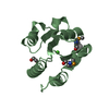
 Corynebacterium glutamicum ATCC 13032 (bacteria)
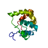
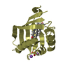
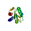
 X-RAY DIFFRACTION /
  Authors
 Citation
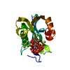
 Structure visualization
 Downloads & links
 Other downloads
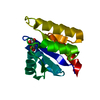


 PDBj
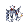
PDBj Assembly
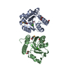
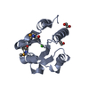

 Mass: 40.078 Da / Num. of mol.: 2 / Source method: obtained synthetically / Formula: Ca
 Mass: 62.068 Da / Num. of mol.: 3 / Source method: obtained synthetically / Formula: C2H6O2
 Mass: 18.015 Da / Num. of mol.: 268 / Source method: isolated from a natural source / Formula: H2O
 Sample preparation
 / Beamline: BL11-1 / Wavelength: 0.91162, 0.97929
 Processing