 Movie
Movie Controller

[English] 日本語
 Yorodumi
- PDB-3dlk: Crystal Structure of an engineered form of the HIV-1 Reverse Tran... -
+ Open data

- Basic information
| Entry | Database: PDB / ID: 3dlk | ||||||
|---|---|---|---|---|---|---|---|
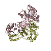
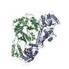
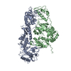
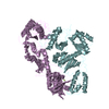
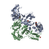
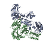
| Title | Crystal Structure of an engineered form of the HIV-1 Reverse Transcriptase, RT69A | ||||||
 Components Components |
| ||||||
 Keywords Keywords | TRANSFERASE / AIDS / HIV / REVERSE TRANSCRIPTASE / RT / crystal engineering / RNA-binding / RNA-directed DNA polymerase / Viral nucleoprotein | ||||||
| Function / homology |  Function and homology information Function and homology informationHIV-1 retropepsin / symbiont-mediated activation of host apoptosis / retroviral ribonuclease H / exoribonuclease H / exoribonuclease H activity / DNA integration / viral genome integration into host DNA / establishment of integrated proviral latency / RNA-directed DNA polymerase / RNA stem-loop binding ...HIV-1 retropepsin / symbiont-mediated activation of host apoptosis / retroviral ribonuclease H / exoribonuclease H / exoribonuclease H activity / DNA integration / viral genome integration into host DNA / establishment of integrated proviral latency / RNA-directed DNA polymerase / RNA stem-loop binding / viral penetration into host nucleus / host multivesicular body / RNA-directed DNA polymerase activity / RNA-DNA hybrid ribonuclease activity / Transferases; Transferring phosphorus-containing groups; Nucleotidyltransferases / host cell / viral nucleocapsid / DNA recombination / DNA-directed DNA polymerase / aspartic-type endopeptidase activity / Hydrolases; Acting on ester bonds / DNA-directed DNA polymerase activity / symbiont-mediated suppression of host gene expression / viral translational frameshifting / symbiont entry into host cell / lipid binding / host cell nucleus / host cell plasma membrane / virion membrane / structural molecule activity / proteolysis / DNA binding / zinc ion binding Similarity search - Function | ||||||
| Biological species |  Human immunodeficiency virus type 1 BH10 Human immunodeficiency virus type 1 BH10 Human immunodeficiency virus type 1 Human immunodeficiency virus type 1 | ||||||
| Method |  X-RAY DIFFRACTION / SYNCHROTRON / Resolution: 1.85 Å X-RAY DIFFRACTION / SYNCHROTRON / Resolution: 1.85 Å | ||||||
 Authors Authors | Ho, W.C. / Bauman, J.D. / Himmel, D.M. / Das, K. / Arnold, E. | ||||||
 Citation Citation | Journal: Nucleic Acids Res. / Year: 2008 Title: Crystal engineering of HIV-1 reverse transcriptase for structure-based drug design. Authors: Bauman, J.D. / Das, K. / Ho, W.C. / Baweja, M. / Himmel, D.M. / Clark, A.D. / Oren, D.A. / Boyer, P.L. / Hughes, S.H. / Shatkin, A.J. / Arnold, E. | ||||||
| History |
|
- Structure visualization
| Structure viewer | Molecule: MolmilJmol/JSmol |
|---|
- Downloads & links
-Download
| PDBx/mmCIF format | 3dlk.cif.gz | 209.2 KB | Display | PDBx/mmCIF format |
|---|---|---|---|---|
| PDB format | pdb3dlk.ent.gz | 165.6 KB | Display | PDB format |
| PDBx/mmJSON format | 3dlk.json.gz | Tree view | PDBx/mmJSON format | |
| Others |  Other downloads Other downloads |
-Validation report
| Arichive directory | https://data.pdbj.org/pub/pdb/validation_reports/dl/3dlkftp://data.pdbj.org/pub/pdb/validation_reports/dl/3dlk | HTTPS FTP |
|---|
-Related structure data
| Related structure data |  2zd1S S: Starting model for refinement |
|---|---|
| Similar structure data |
-Links
 PDBj
PDBj


- Assembly
| Deposited unit | 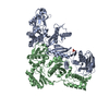
| ||||||||
|---|---|---|---|---|---|---|---|---|---|
| 1 |
| ||||||||
| Unit cell |
|
-Components
| #1: Protein | Mass: 63946.211 Da / Num. of mol.: 1 / Fragment: p66 (UNP residues 599 to 1153) / Mutation: K172A, K173A, C258Q Source method: isolated from a genetically manipulated source Source: (gene. exp.) Human immunodeficiency virus type 1 BH10Strain: ISOLATE BH10 / Gene: gag-pol / Production host:  References: UniProt: P03366, RNA-directed DNA polymerase, DNA-directed DNA polymerase |
|---|---|
| #2: Protein | Mass: 49531.871 Da / Num. of mol.: 1 / Fragment: p51 (UNP residues 605 to 1027) Source method: isolated from a genetically manipulated source Source: (gene. exp.) Human immunodeficiency virus type 1 / Gene: gag-pol / Production host: References: UniProt: P03366, DNA-directed DNA polymerase, ribonuclease H |
| #3: Chemical | ChemComp-SO4 /   Mass: 96.063 Da / Num. of mol.: 1 / Source method: obtained synthetically / Formula: SO4 Mass: 96.063 Da / Num. of mol.: 1 / Source method: obtained synthetically / Formula: SO4 |
| #4: Water | ChemComp-HOH /  Mass: 18.015 Da / Num. of mol.: 187 / Source method: isolated from a natural source / Formula: H2O Mass: 18.015 Da / Num. of mol.: 187 / Source method: isolated from a natural source / Formula: H2O |
-Experimental details
-Experiment
| Experiment | Method: X-RAY DIFFRACTION / Number of used crystals: 1 |
|---|
- Sample preparation
| Crystal | Density Matthews: 2.77 Å3/Da / Density % sol: 55.54 % |
|---|---|
| Crystal grow | Temperature: 279.15 K / Method: vapor diffusion / Details: vapor diffusion, temperature 279.15K |
-Data collection
| Diffraction | Mean temperature: 93.15 K |
|---|---|
| Diffraction source | Source: SYNCHROTRON / Site: CHESS  / Beamline: F1 / Beamline: F1 |
| Detector | Type: ADSC QUANTUM 270 / Detector: CCD |
| Radiation | Protocol: SINGLE WAVELENGTH / Monochromatic (M) / Laue (L): M / Scattering type: x-ray |
| Radiation wavelength | Relative weight: 1 |
| Reflection | Resolution: 1.85→40.66 Å / Num. obs: 99442 |
- Processing
| Software |
| ||||||||||||||||||||
|---|---|---|---|---|---|---|---|---|---|---|---|---|---|---|---|---|---|---|---|---|---|
| Refinement | Starting model: PDB entry 2zd1 Resolution: 1.85→40.66 Å / Occupancy max: 1 / Occupancy min: 1 / σ(F): 0
| ||||||||||||||||||||
| Solvent computation | Bsol: 57.805 Å2 | ||||||||||||||||||||
| Displacement parameters | Biso max: 150.19 Å2 / Biso mean: 50.143 Å2 / Biso min: 0.11 Å2
| ||||||||||||||||||||
| Refinement step | Cycle: LAST / Resolution: 1.85→40.66 Å
| ||||||||||||||||||||
| Refine LS restraints |
| ||||||||||||||||||||
| Xplor file |
|
