 Movie
Movie Controller

[English] 日本語
 Yorodumi
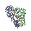
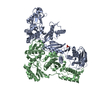
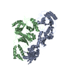
- PDB-3ig1: HIV-1 Reverse Transcriptase with the Inhibitor beta-Thujaplicinol... -
+ Open data

- Basic information
| Entry | Database: PDB / ID: 3ig1 | ||||||
|---|---|---|---|---|---|---|---|
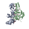
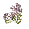
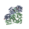
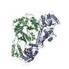
| Title | HIV-1 Reverse Transcriptase with the Inhibitor beta-Thujaplicinol Bound at the RNase H Active Site | ||||||
 Components Components |
| ||||||
 Keywords Keywords | TRANSFERASE / RNASE H INHIBITOR / PROTEIN-INHIBITOR COMPLEX / STRUCTURE-BASED DRUG DESIGN / TROPOLONES / TROPYLIUM ION / DIVALENT CATION CHELATOR / AIDS / DNA-DIRECTED DNA POLYMERASE / METAL-BINDING / MULTIFUNCTIONAL ENZYME / NUCLEOTIDYLTRANSFERASE / RNA-BINDING / RNA-DIRECTED DNA POLYMERASE | ||||||
| Function / homology |  Function and homology information Function and homology informationHIV-1 retropepsin / symbiont-mediated activation of host apoptosis / retroviral ribonuclease H / exoribonuclease H / exoribonuclease H activity / DNA integration / viral genome integration into host DNA / establishment of integrated proviral latency / RNA-directed DNA polymerase / RNA stem-loop binding ...HIV-1 retropepsin / symbiont-mediated activation of host apoptosis / retroviral ribonuclease H / exoribonuclease H / exoribonuclease H activity / DNA integration / viral genome integration into host DNA / establishment of integrated proviral latency / RNA-directed DNA polymerase / RNA stem-loop binding / viral penetration into host nucleus / host multivesicular body / RNA-directed DNA polymerase activity / RNA-DNA hybrid ribonuclease activity / Transferases; Transferring phosphorus-containing groups; Nucleotidyltransferases / host cell / viral nucleocapsid / DNA recombination / DNA-directed DNA polymerase / aspartic-type endopeptidase activity / Hydrolases; Acting on ester bonds / DNA-directed DNA polymerase activity / symbiont-mediated suppression of host gene expression / viral translational frameshifting / symbiont entry into host cell / lipid binding / host cell nucleus / host cell plasma membrane / virion membrane / structural molecule activity / proteolysis / DNA binding / zinc ion binding Similarity search - Function | ||||||
| Biological species |  Human immunodeficiency virus type 1 BH10 Human immunodeficiency virus type 1 BH10 | ||||||
| Method |  X-RAY DIFFRACTION / SYNCHROTRON / MOLECULAR REPLACEMENT / molecular replacement / Resolution: 2.8 Å X-RAY DIFFRACTION / SYNCHROTRON / MOLECULAR REPLACEMENT / molecular replacement / Resolution: 2.8 Å | ||||||
 Authors Authors | Himmel, D.M. / Maegley, K.A. / Pauly, T.A. / Arnold, E. | ||||||
 Citation Citation | Journal: Structure / Year: 2009 Title: Structure of HIV-1 reverse transcriptase with the inhibitor beta-Thujaplicinol bound at the RNase H active site. Authors: Himmel, D.M. / Maegley, K.A. / Pauly, T.A. / Bauman, J.D. / Das, K. / Dharia, C. / Clark, A.D. / Ryan, K. / Hickey, M.J. / Love, R.A. / Hughes, S.H. / Bergqvist, S. / Arnold, E. #1: Journal: Nucleic Acids Res. / Year: 2008Title: Crystal Engineering of HIV-1 Reverse Transcriptase for Structure-based Drug Design Authors: Bauman, J.D. / Das, K. / Ho, W.C. / Baweja, M. / Himmel, D.M. / Clark Jr., A.D. / Oren, D.A. / Boyer, P.L. / Hughes, S.H. / Shatkin, A.J. / Arnold, E. | ||||||
| History |
|
- Structure visualization
| Structure viewer | Molecule: MolmilJmol/JSmol |
|---|
- Downloads & links
-Download
| PDBx/mmCIF format | 3ig1.cif.gz | 209.2 KB | Display | PDBx/mmCIF format |
|---|---|---|---|---|
| PDB format | pdb3ig1.ent.gz | 165.7 KB | Display | PDB format |
| PDBx/mmJSON format | 3ig1.json.gz | Tree view | PDBx/mmJSON format | |
| Others |  Other downloads Other downloads |
-Validation report
| Arichive directory | https://data.pdbj.org/pub/pdb/validation_reports/ig/3ig1ftp://data.pdbj.org/pub/pdb/validation_reports/ig/3ig1 | HTTPS FTP |
|---|
-Related structure data
| Related structure data |  3k2pC 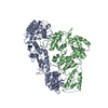 1dloS S: Starting model for refinement C: citing same article ( |
|---|---|
| Similar structure data |


-Links
 PDBj
PDBj


- Assembly
| Deposited unit | 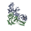
| ||||||||
|---|---|---|---|---|---|---|---|---|---|
| 1 |
| ||||||||
| Unit cell |
|
-Components
| #1: Protein | Mass: 63815.016 Da / Num. of mol.: 1 / Fragment: p66 subunit, residues 600-1154 / Mutation: F759S, C879S Source method: isolated from a genetically manipulated source Details: See citation_id 1 above. Source: (gene. exp.) Human immunodeficiency virus type 1 BH10Gene: gag-pol, POL / Plasmid: pCDF-2 / Production host:  | ||
|---|---|---|---|
| #2: Protein | Mass: 50039.488 Da / Num. of mol.: 1 / Fragment: p51 subunit, residues 600-1027 / Mutation: C879S Source method: isolated from a genetically manipulated source Details: See citation_id 1 above. Source: (gene. exp.) Human immunodeficiency virus type 1 BH10Gene: gag-pol, POL / Plasmid: pCDF-2 / Production host: | ||
| #3: Chemical | ChemComp-JTH / 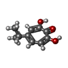  Mass: 180.200 Da / Num. of mol.: 1 / Source method: obtained synthetically / Formula: C10H12O3 Mass: 180.200 Da / Num. of mol.: 1 / Source method: obtained synthetically / Formula: C10H12O3 | ||
| #4: Chemical |   Mass: 54.938 Da / Num. of mol.: 2 / Source method: obtained synthetically / Formula: Mn Mass: 54.938 Da / Num. of mol.: 2 / Source method: obtained synthetically / Formula: Mn#5: Water | ChemComp-HOH / |  Mass: 18.015 Da / Num. of mol.: 74 / Source method: isolated from a natural source / Formula: H2O Mass: 18.015 Da / Num. of mol.: 74 / Source method: isolated from a natural source / Formula: H2O |
-Experimental details
-Experiment
| Experiment | Method: X-RAY DIFFRACTION / Number of used crystals: 1 |
|---|
- Sample preparation
| Crystal | Density Matthews: 2.69 Å3/Da / Density % sol: 54.3 % |
|---|---|
| Crystal grow | Temperature: 277 K / Method: vapor diffusion, hanging drop / pH: 8.2 Details: 50 mM Bicine pH 8.2, 45 mM Ammonium Sulfate, 15 mM Manganese Sulfate, 10 mM Spermine, 5 mM Taurine, 2% PEG 400, 10% PEG 8000 Combined with equal volume of: 10 mM Tris pH 8.0, 75 mM NaCl, 3. ...Details: 50 mM Bicine pH 8.2, 45 mM Ammonium Sulfate, 15 mM Manganese Sulfate, 10 mM Spermine, 5 mM Taurine, 2% PEG 400, 10% PEG 8000 Combined with equal volume of: 10 mM Tris pH 8.0, 75 mM NaCl, 3.5% DMSO, 0.86 mM b-Thujaplicinol, 0.17 mM Reverse Transcriptase, VAPOR DIFFUSION, HANGING DROP, temperature 277K |
-Data collection
| Diffraction | Mean temperature: 100 K |
|---|---|
| Diffraction source | Source: SYNCHROTRON / Site: NSLS  / Beamline: X25 / Wavelength: 1.08 Å / Beamline: X25 / Wavelength: 1.08 Å |
| Detector | Type: ADSC QUANTUM 315 / Detector: CCD / Date: Sep 20, 2007 |
| Radiation | Monochromator: Double silicon(111) crystal monochromator and mirror Protocol: SINGLE WAVELENGTH / Monochromatic (M) / Laue (L): M / Scattering type: x-ray |
| Radiation wavelength | Wavelength: 1.08 Å / Relative weight: 1 |
| Reflection | Resolution: 2.8→35 Å / Num. all: 28769 / Num. obs: 28769 / % possible obs: 94.3 % / Observed criterion σ(I): -1.1 / Redundancy: 2.7 % / Biso Wilson estimate: 71.5 Å2 / Rsym value: 0.061 / Χ2: 1.006 / Net I/σ(I): 15.3 |
| Reflection shell | Resolution: 2.8→2.9 Å / Redundancy: 2.1 % / Num. unique all: 2599 / Rsym value: 0.489 / Χ2: 1.005 / % possible all: 77.2 |
-Phasing
| Phasing | Method: molecular replacement |
|---|
- Processing
| Software |
| ||||||||||||||||||||||||||||||||||||
|---|---|---|---|---|---|---|---|---|---|---|---|---|---|---|---|---|---|---|---|---|---|---|---|---|---|---|---|---|---|---|---|---|---|---|---|---|---|
| Refinement | Method to determine structure: MOLECULAR REPLACEMENT Starting model: PDB ENTRY 1DLO Resolution: 2.8→33.75 Å / Rfactor Rfree error: 0.009 / Occupancy max: 1 / Occupancy min: 1 / FOM work R set: 0.791 / Data cutoff high absF: 199160 / Data cutoff low absF: 0 / Isotropic thermal model: RESTRAINED / Cross valid method: THROUGHOUT / σ(F): 0 / Stereochemistry target values: Engh & Huber
| ||||||||||||||||||||||||||||||||||||
| Solvent computation | Solvent model: FLAT MODEL / Bsol: 52.5 Å2 / ksol: 0.297 e/Å3 | ||||||||||||||||||||||||||||||||||||
| Displacement parameters | Biso max: 181.49 Å2 / Biso mean: 89.018 Å2 / Biso min: 26.04 Å2
| ||||||||||||||||||||||||||||||||||||
| Refine analyze |
| ||||||||||||||||||||||||||||||||||||
| Refinement step | Cycle: LAST / Resolution: 2.8→33.75 Å
| ||||||||||||||||||||||||||||||||||||
| Refine LS restraints |
| ||||||||||||||||||||||||||||||||||||
| LS refinement shell | Resolution: 2.8→2.98 Å / Rfactor Rfree error: 0.044 / Total num. of bins used: 6
| ||||||||||||||||||||||||||||||||||||
| Xplor file |
|
