 Movie
Movie Controller

[English] 日本語
 Yorodumi
- PDB-3d2s: Crystal structure of MBNL1 tandem zinc finger 3 and 4 domain in c... -
+ Open data

- Basic information
| Entry | Database: PDB / ID: 3d2s | ||||||
|---|---|---|---|---|---|---|---|
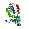
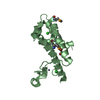
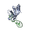
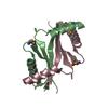
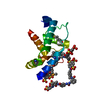
| Title | Crystal structure of MBNL1 tandem zinc finger 3 and 4 domain in complex with CGCUGU RNA | ||||||
 Components Components |
| ||||||
 Keywords Keywords | METAL BINDING PROTEIN/RNA / tandem zinc finger domain / RNA / Metal-binding / Nucleus / RNA-binding / Zinc-finger / METAL BINDING PROTEIN-RNA COMPLEX | ||||||
| Function / homology |  Function and homology information Function and homology informationmyoblast differentiation / embryonic limb morphogenesis / regulation of RNA splicing / RNA splicing / mRNA processing / cytoplasmic stress granule / nervous system development / double-stranded RNA binding / in utero embryonic development / RNA binding ...myoblast differentiation / embryonic limb morphogenesis / regulation of RNA splicing / RNA splicing / mRNA processing / cytoplasmic stress granule / nervous system development / double-stranded RNA binding / in utero embryonic development / RNA binding / zinc ion binding / nucleoplasm / nucleus / cytoplasm / cytosol Similarity search - Function | ||||||
| Biological species |  Homo sapiens (human) Homo sapiens (human)Synthetic construct (others) | ||||||
| Method |  X-RAY DIFFRACTION / SYNCHROTRON / MOLECULAR REPLACEMENT / Resolution: 1.7 Å X-RAY DIFFRACTION / SYNCHROTRON / MOLECULAR REPLACEMENT / Resolution: 1.7 Å | ||||||
 Authors Authors | Teplova, M. / Patel, D.J. | ||||||
 Citation Citation | Journal: Nat.Struct.Mol.Biol. / Year: 2008 Title: Structural insights into RNA recognition by the alternative-splicing regulator muscleblind-like MBNL1. Authors: Teplova, M. / Patel, D.J. | ||||||
| History |
|
- Structure visualization
| Structure viewer | Molecule: MolmilJmol/JSmol |
|---|
- Downloads & links
-Download
| PDBx/mmCIF format | 3d2s.cif.gz | 87.5 KB | Display | PDBx/mmCIF format |
|---|---|---|---|---|
| PDB format | pdb3d2s.ent.gz | 65.3 KB | Display | PDB format |
| PDBx/mmJSON format | 3d2s.json.gz | Tree view | PDBx/mmJSON format | |
| Others |  Other downloads Other downloads |
-Validation report
| Arichive directory | https://data.pdbj.org/pub/pdb/validation_reports/d2/3d2sftp://data.pdbj.org/pub/pdb/validation_reports/d2/3d2s | HTTPS FTP |
|---|
-Related structure data


-Links
 PDBj
PDBj
- Assembly
| Deposited unit | 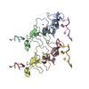
| ||||||||
|---|---|---|---|---|---|---|---|---|---|
| 1 | 
| ||||||||
| 2 | 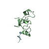
| ||||||||
| 3 | 
| ||||||||
| 4 | 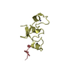
| ||||||||
| Unit cell |
|
-Components
| #1: RNA chain | Mass: 1868.149 Da / Num. of mol.: 4 / Source method: obtained synthetically / Details: Chemically synthesized RNA oligonucleotide / Source: (synth.) Synthetic construct (others) #2: Protein | Mass: 8169.182 Da / Num. of mol.: 4 Fragment: tandem zinc finger 3 and 4 domains (UNP residues 178-246) Source method: isolated from a genetically manipulated source Source: (gene. exp.) Homo sapiens (human) / Gene: MBNL1, EXP, KIAA0428, MBNL / Plasmid: Pet28b / Production host:  #3: Chemical | ChemComp-ZN /   Mass: 65.409 Da / Num. of mol.: 8 / Source method: obtained synthetically / Formula: Zn Mass: 65.409 Da / Num. of mol.: 8 / Source method: obtained synthetically / Formula: Zn#4: Water | ChemComp-HOH / |  Mass: 18.015 Da / Num. of mol.: 402 / Source method: isolated from a natural source / Formula: H2O Mass: 18.015 Da / Num. of mol.: 402 / Source method: isolated from a natural source / Formula: H2O |
|---|
-Experimental details
-Experiment
| Experiment | Method: X-RAY DIFFRACTION / Number of used crystals: 1 |
|---|
- Sample preparation
| Crystal | Density Matthews: 2.2 Å3/Da / Density % sol: 44.18 % | ||||||||||||||||||||||||||||
|---|---|---|---|---|---|---|---|---|---|---|---|---|---|---|---|---|---|---|---|---|---|---|---|---|---|---|---|---|---|
| Crystal grow | Temperature: 277 K / Method: vapor diffusion / pH: 5.5 Details: 25% PEG 3350, 0.1M Bis-Tris, pH 5.5, VAPOR DIFFUSION, temperature 277K | ||||||||||||||||||||||||||||
| Components of the solutions |
|
-Data collection
| Diffraction | Mean temperature: 100 K |
|---|---|
| Diffraction source | Source: SYNCHROTRON / Site: APS  / Beamline: 24-ID-C / Wavelength: 0.9795 Å / Beamline: 24-ID-C / Wavelength: 0.9795 Å |
| Detector | Type: ADSC QUANTUM 315 / Detector: CCD / Date: Dec 20, 2006 |
| Radiation | Protocol: SINGLE WAVELENGTH / Monochromatic (M) / Laue (L): M / Scattering type: x-ray |
| Radiation wavelength | Wavelength: 0.9795 Å / Relative weight: 1 |
| Reflection | Resolution: 1.7→50 Å / Num. obs: 38651 / % possible obs: 99.9 % / Observed criterion σ(I): 2 / Rsym value: 0.074 / Net I/σ(I): 17.7 |
| Reflection shell | Resolution: 1.7→1.76 Å / Redundancy: 4.8 % / Mean I/σ(I) obs: 3.6 / Num. unique all: 3846 / Rsym value: 0.44 / % possible all: 100 |
- Processing
| Software |
| ||||||||||||||||||||||||||||||||||||||||||||||||||||||||||||||||||||||||||||||||||||||||||||||||||||||||||||||||||||||||||||||||||||||||||||||||||||||||||||||||||||||||||
|---|---|---|---|---|---|---|---|---|---|---|---|---|---|---|---|---|---|---|---|---|---|---|---|---|---|---|---|---|---|---|---|---|---|---|---|---|---|---|---|---|---|---|---|---|---|---|---|---|---|---|---|---|---|---|---|---|---|---|---|---|---|---|---|---|---|---|---|---|---|---|---|---|---|---|---|---|---|---|---|---|---|---|---|---|---|---|---|---|---|---|---|---|---|---|---|---|---|---|---|---|---|---|---|---|---|---|---|---|---|---|---|---|---|---|---|---|---|---|---|---|---|---|---|---|---|---|---|---|---|---|---|---|---|---|---|---|---|---|---|---|---|---|---|---|---|---|---|---|---|---|---|---|---|---|---|---|---|---|---|---|---|---|---|---|---|---|---|---|---|---|---|
| Refinement | Method to determine structure: MOLECULAR REPLACEMENT Starting model: MBNL1 ZnF3/4 structure Resolution: 1.7→20 Å / Cor.coef. Fo:Fc: 0.964 / Cor.coef. Fo:Fc free: 0.949 / SU B: 5.62 / SU ML: 0.095 / TLS residual ADP flag: LIKELY RESIDUAL / Cross valid method: THROUGHOUT / ESU R: 0.134 / ESU R Free: 0.128 / Stereochemistry target values: MAXIMUM LIKELIHOOD / Details: HYDROGENS HAVE BEEN ADDED IN THE RIDING POSITIONS
| ||||||||||||||||||||||||||||||||||||||||||||||||||||||||||||||||||||||||||||||||||||||||||||||||||||||||||||||||||||||||||||||||||||||||||||||||||||||||||||||||||||||||||
| Solvent computation | Ion probe radii: 0.8 Å / Shrinkage radii: 0.8 Å / VDW probe radii: 1.2 Å / Solvent model: MASK | ||||||||||||||||||||||||||||||||||||||||||||||||||||||||||||||||||||||||||||||||||||||||||||||||||||||||||||||||||||||||||||||||||||||||||||||||||||||||||||||||||||||||||
| Displacement parameters | Biso mean: 14.101 Å2
| ||||||||||||||||||||||||||||||||||||||||||||||||||||||||||||||||||||||||||||||||||||||||||||||||||||||||||||||||||||||||||||||||||||||||||||||||||||||||||||||||||||||||||
| Refinement step | Cycle: LAST / Resolution: 1.7→20 Å
| ||||||||||||||||||||||||||||||||||||||||||||||||||||||||||||||||||||||||||||||||||||||||||||||||||||||||||||||||||||||||||||||||||||||||||||||||||||||||||||||||||||||||||
| Refine LS restraints |
| ||||||||||||||||||||||||||||||||||||||||||||||||||||||||||||||||||||||||||||||||||||||||||||||||||||||||||||||||||||||||||||||||||||||||||||||||||||||||||||||||||||||||||
| LS refinement shell | Resolution: 1.698→1.742 Å / Total num. of bins used: 20
| ||||||||||||||||||||||||||||||||||||||||||||||||||||||||||||||||||||||||||||||||||||||||||||||||||||||||||||||||||||||||||||||||||||||||||||||||||||||||||||||||||||||||||
| Refinement TLS params. | Method: refined / Origin x: 13.995 Å / Origin y: 3.97 Å / Origin z: 13.604 Å
| ||||||||||||||||||||||||||||||||||||||||||||||||||||||||||||||||||||||||||||||||||||||||||||||||||||||||||||||||||||||||||||||||||||||||||||||||||||||||||||||||||||||||||
| Refinement TLS group |
|
