Resolution: 2.1→2.18 Å / Redundancy: 2.1 % / Rmerge(I) obs: 0.666 / Mean I/σ(I) obs: 1.3 / Num. unique all: 4027 / % possible all: 96.8
-
Processing
Software
Name
Version
Classification
NB
PHENIX
1.7.2_869
refinement
PDB_EXTRACT
3.1
dataextraction
ADSC
Quantum
datacollection
HKL-2000
datareduction
HKL-2000
datascaling
SHELXS
phasing
Refinement
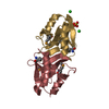
Method to determine structure: SAD / Resolution: 2.099→43.348 Å / Occupancy max: 1 / Occupancy min: 0.22 / SU ML: 0.68 / Cross valid method: THROUGHOUT / σ(F): 1.05 / Phase error: 25.7 / Stereochemistry target values: ML Details: CYS108 SIDE CHAIN IS COVALENTLY MODIFIED BASED ON THE ELECTRON DENSITY. HOWEVER, THE IDENTITY OF THE MODIFYING GROUP IS NOT KNOWN.THE EXTRA ELECTRON DENSITY IS CURRENTLY MODELED BY WATERS 605, 640, 668 AND 669.
Rfactor
Num. reflection
% reflection
Selection details
Rfree
0.249
2172
5.14 %
RANDOM
Rwork
0.214
-
-
-
obs
0.216
42280
99.35 %
-
Solvent computation
Shrinkage radii: 0.9 Å / VDW probe radii: 1.11 Å / Solvent model: FLAT BULK SOLVENT MODEL / Bsol: 44.938 Å2 / ksol: 0.329 e/Å3
In the structure databanks used in Yorodumi, some data are registered as the other names, "COVID-19 virus" and "2019-nCoV". Here are the details of the virus and the list of structure data.
Jan 31, 2019. EMDB accession codes are about to change! (news from PDBe EMDB page)
EMDB accession codes are about to change! (news from PDBe EMDB page)
The allocation of 4 digits for EMDB accession codes will soon come to an end. Whilst these codes will remain in use, new EMDB accession codes will include an additional digit and will expand incrementally as the available range of codes is exhausted. The current 4-digit format prefixed with “EMD-” (i.e. EMD-XXXX) will advance to a 5-digit format (i.e. EMD-XXXXX), and so on. It is currently estimated that the 4-digit codes will be depleted around Spring 2019, at which point the 5-digit format will come into force.
The EM Navigator/Yorodumi systems omit the EMD- prefix.
Related info.:Q: What is EMD? / ID/Accession-code notation in Yorodumi/EM Navigator
Yorodumi is a browser for structure data from EMDB, PDB, SASBDB, etc.
This page is also the successor to EM Navigator detail page, and also detail information page/front-end page for Omokage search.
The word "yorodu" (or yorozu) is an old Japanese word meaning "ten thousand". "mi" (miru) is to see.
Related info.:EMDB / PDB / SASBDB / Comparison of 3 databanks / Yorodumi Search / Aug 31, 2016. New EM Navigator & Yorodumi / Yorodumi Papers / Jmol/JSmol / Function and homology information / Changes in new EM Navigator and Yorodumi
 Movie
Movie Controller

 Yorodumi
 Open data
 Basic information
 Components
 Keywords
 Function and homology information
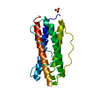
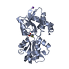
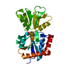
 Enterobacteria phage lambda (virus)
 X-RAY DIFFRACTION /
  Authors
 Citation
 Structure visualization
 Downloads & links
 Other downloads


 PDBj
PDBj

 Assembly
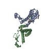

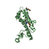


 Mass: 65.409 Da / Num. of mol.: 2 / Source method: obtained synthetically / Formula: Zn
 Mass: 79.904 Da / Num. of mol.: 3 / Source method: obtained synthetically / Formula: Br
 Mass: 35.453 Da / Num. of mol.: 3 / Source method: obtained synthetically / Formula: Cl
 Mass: 18.015 Da / Num. of mol.: 162 / Source method: isolated from a natural source / Formula: H2O
 Sample preparation
 / Beamline: X4A / Wavelength: 0.97931 Å
 Processing