 Movie
Movie Controller

[English] 日本語
 Yorodumi
- PDB-1z0p: Crystal structure of the Protein of Unknown Function SPY1572 from... -
+ Open data

- Basic information
| Entry | Database: PDB / ID: 1z0p | ||||||
|---|---|---|---|---|---|---|---|
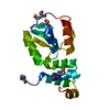
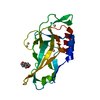
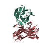
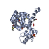
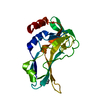
| Title | Crystal structure of the Protein of Unknown Function SPY1572 from Streptococcus pyogenes | ||||||
 Components Components | hypothetical protein SPy1572 | ||||||
 Keywords Keywords | STRUCTURAL GENOMICS / UNKNOWN FUNCTION / Streptococcus pyogenes / PSI / Protein Structure Initiative / Midwest Center for Structural Genomics / MCSG | ||||||
| Function / homology | Protein of unknown function DUF1912 / SPy1572-like superfamily / Domain of unknown function (DUF1912) / Methane Monooxygenase Hydroxylase; Chain G, domain 1 - #90 / Methane Monooxygenase Hydroxylase; Chain G, domain 1 / Up-down Bundle / Mainly Alpha / Aldose 1-epimerase Function and homology information Function and homology information | ||||||
| Biological species |  Streptococcus pyogenes (bacteria) Streptococcus pyogenes (bacteria) | ||||||
| Method |  X-RAY DIFFRACTION / SYNCHROTRON / SAD / Resolution: 1.7 Å X-RAY DIFFRACTION / SYNCHROTRON / SAD / Resolution: 1.7 Å | ||||||
 Authors Authors | Zhang, R. / Lezondra, L. / Clancy, S. / Collart, F. / Joachimiak, A. / Midwest Center for Structural Genomics (MCSG) | ||||||
 Citation Citation | Journal: To be Published Title: The 1.7A Crystal structure of the hypothetical protein SPy1572 from Streptococcus pyogenes Authors: Zhang, R. / Lezondra, L. / Clancy, S. / Collart, F. / Joachimiak, A. | ||||||
| History |
|
- Structure visualization
| Structure viewer | Molecule: MolmilJmol/JSmol |
|---|
- Downloads & links
-Download
| PDBx/mmCIF format | 1z0p.cif.gz | 28 KB | Display | PDBx/mmCIF format |
|---|---|---|---|---|
| PDB format | pdb1z0p.ent.gz | 18.2 KB | Display | PDB format |
| PDBx/mmJSON format | 1z0p.json.gz | Tree view | PDBx/mmJSON format | |
| Others |  Other downloads Other downloads |
-Validation report
| Arichive directory | https://data.pdbj.org/pub/pdb/validation_reports/z0/1z0pftp://data.pdbj.org/pub/pdb/validation_reports/z0/1z0p | HTTPS FTP |
|---|
-Related structure data
| Similar structure data | |
|---|---|
| Other databases |


-Links
 PDBj
PDBj- Assembly
| Deposited unit | 
| ||||||||
|---|---|---|---|---|---|---|---|---|---|
| 1 | 
| ||||||||
| Unit cell |
| ||||||||
| Details | This protein existed as dimer.The second part of the biological assembly is generated by the axis: -x+y, -x, z |
-Components
| #1: Protein | Mass: 9947.979 Da / Num. of mol.: 1 Source method: isolated from a genetically manipulated source Source: (gene. exp.) Streptococcus pyogenes (bacteria) / Strain: M1 GAS / Gene: gi:13622656 / Plasmid: pET15b / Species (production host): Escherichia coli / Production host: |
|---|---|
| #2: Water | ChemComp-HOH /  Mass: 18.015 Da / Num. of mol.: 66 / Source method: isolated from a natural source / Formula: H2O Mass: 18.015 Da / Num. of mol.: 66 / Source method: isolated from a natural source / Formula: H2O |
-Experimental details
-Experiment
| Experiment | Method: X-RAY DIFFRACTION / Number of used crystals: 1 |
|---|
- Sample preparation
| Crystal | Density Matthews: 1.885 Å3/Da / Density % sol: 32.23 % |
|---|---|
| Crystal grow | Temperature: 298 K / Method: vapor diffusion, sitting drop / pH: 4.5 Details: 20% butanediol, 0.1M acetate, pH 4.5, VAPOR DIFFUSION, SITTING DROP, temperature 298K |
-Data collection
| Diffraction | Mean temperature: 100 K |
|---|---|
| Diffraction source | Source: SYNCHROTRON / Site: APS  / Beamline: 19-ID / Wavelength: 0.9795 Å / Beamline: 19-ID / Wavelength: 0.9795 Å |
| Detector | Type: ADSC QUANTUM 4 / Detector: CCD / Date: Dec 4, 2004 / Details: mirrors |
| Radiation | Monochromator: Si 111 channel / Protocol: SINGLE WAVELENGTH / Monochromatic (M) / Laue (L): M / Scattering type: x-ray |
| Radiation wavelength | Wavelength: 0.9795 Å / Relative weight: 1 |
| Reflection | Resolution: 1.7→40 Å / Num. obs: 16058 / % possible obs: 99.7 % / Observed criterion σ(F): 2 / Observed criterion σ(I): 2 / Redundancy: 10.1 % / Biso Wilson estimate: 27 Å2 / Rmerge(I) obs: 0.067 / Net I/σ(I): 41.88 |
| Reflection shell | Resolution: 1.7→1.76 Å / Redundancy: 8.2 % / Rmerge(I) obs: 0.596 / Mean I/σ(I) obs: 2.83 / Num. unique all: 828 / % possible all: 99.6 |
- Processing
| Software |
| |||||||||||||||||||||||||
|---|---|---|---|---|---|---|---|---|---|---|---|---|---|---|---|---|---|---|---|---|---|---|---|---|---|---|
| Refinement | Method to determine structure: SAD / Resolution: 1.7→25.69 Å / Rfactor Rfree error: 0.009 / Data cutoff high absF: 481465.31 / Data cutoff low absF: 0 / Isotropic thermal model: RESTRAINED / Cross valid method: THROUGHOUT / σ(F): 0 / Stereochemistry target values: Engh & Huber
| |||||||||||||||||||||||||
| Solvent computation | Solvent model: FLAT MODEL / Bsol: 81.5796 Å2 / ksol: 0.382325 e/Å3 | |||||||||||||||||||||||||
| Displacement parameters | Biso mean: 32.5 Å2
| |||||||||||||||||||||||||
| Refine analyze |
| |||||||||||||||||||||||||
| Refinement step | Cycle: LAST / Resolution: 1.7→25.69 Å
| |||||||||||||||||||||||||
| Refine LS restraints |
| |||||||||||||||||||||||||
| LS refinement shell | Resolution: 1.7→1.81 Å / Rfactor Rfree error: 0.031 / Total num. of bins used: 6
| |||||||||||||||||||||||||
| Xplor file |
|
