 Movie
Movie Controller

[English] 日本語
 Yorodumi
- PDB-3cyo: Structure of a longer thermalstable core domain of HIV-1 GP41 con... -
+ Open data

- Basic information
| Entry | Database: PDB / ID: 3cyo | ||||||
|---|---|---|---|---|---|---|---|
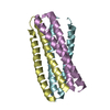
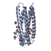
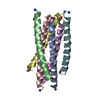
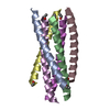
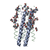
| Title | Structure of a longer thermalstable core domain of HIV-1 GP41 containing the enfuvirtide resistance mutation N43D and complementary mutation E137K | ||||||
 Components Components | Transmembrane protein | ||||||
 Keywords Keywords | VIRAL PROTEIN / HIV-1 ENVELOPE GLYCOPROTEIN / 6-HELIX BUNDLE / GP41 / N43D / E137K / AIDS / APOPTOSIS / COILED COIL / ENVELOPE PROTEIN / FUSION PROTEIN / HOST-VIRUS INTERACTION / MEMBRANE / TRANSMEMBRANE / VIRION / Cleavage on pair of basic residues / Lipoprotein / Palmitate / Viral immunoevasion | ||||||
| Function / homology |  Function and homology information Function and homology informationmembrane fusion involved in viral entry into host cell / Dectin-2 family / symbiont-mediated perturbation of host defense response / positive regulation of plasma membrane raft polarization / positive regulation of receptor clustering / host cell endosome membrane / clathrin-dependent endocytosis of virus by host cell / viral protein processing / fusion of virus membrane with host plasma membrane / fusion of virus membrane with host endosome membrane ...membrane fusion involved in viral entry into host cell / Dectin-2 family / symbiont-mediated perturbation of host defense response / positive regulation of plasma membrane raft polarization / positive regulation of receptor clustering / host cell endosome membrane / clathrin-dependent endocytosis of virus by host cell / viral protein processing / fusion of virus membrane with host plasma membrane / fusion of virus membrane with host endosome membrane / viral envelope / symbiont entry into host cell / virion attachment to host cell / host cell plasma membrane / virion membrane / structural molecule activity / membrane Similarity search - Function | ||||||
| Biological species |   Human immunodeficiency virus type 1 Human immunodeficiency virus type 1 | ||||||
| Method |  X-RAY DIFFRACTION / MOLECULAR REPLACEMENT / Resolution: 2.1 Å X-RAY DIFFRACTION / MOLECULAR REPLACEMENT / Resolution: 2.1 Å | ||||||
 Authors Authors | Wang, Z.M. / Dwyer, J.J. | ||||||
 Citation Citation | Journal: Biochemistry / Year: 2008 Title: Impact of the enfuvirtide resistance mutation N43D and the associated baseline polymorphism E137K on peptide sensitivity and six-helix bundle structure. Authors: Bai, X. / Wilson, K.L. / Seedorff, J.E. / Ahrens, D. / Green, J. / Davison, D.K. / Jin, L. / Stanfield-Oakley, S.A. / Mosier, S.M. / Melby, T.E. / Cammack, N. / Wang, Z. / Greenberg, M.L. / Dwyer, J.J. | ||||||
| History |
|
- Structure visualization
| Structure viewer | Molecule: MolmilJmol/JSmol |
|---|
- Downloads & links
-Download
| PDBx/mmCIF format | 3cyo.cif.gz | 27 KB | Display | PDBx/mmCIF format |
|---|---|---|---|---|
| PDB format | pdb3cyo.ent.gz | 17.7 KB | Display | PDB format |
| PDBx/mmJSON format | 3cyo.json.gz | Tree view | PDBx/mmJSON format | |
| Others |  Other downloads Other downloads |
-Validation report
| Arichive directory | https://data.pdbj.org/pub/pdb/validation_reports/cy/3cyoftp://data.pdbj.org/pub/pdb/validation_reports/cy/3cyo | HTTPS FTP |
|---|
-Related structure data
| Related structure data |  2ot5C  3cp1C  1envS C: citing same article ( S: Starting model for refinement |
|---|---|
| Similar structure data |


-Links
 PDBj
PDBj


- Assembly
| Deposited unit | 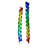
| |||||||||||||||
|---|---|---|---|---|---|---|---|---|---|---|---|---|---|---|---|---|
| 1 | 
| |||||||||||||||
| Unit cell |
| |||||||||||||||
| Components on special symmetry positions |
|
-Components
| #1: Protein | Mass: 9975.190 Da / Num. of mol.: 1 Fragment: FUSION PROTEIN OF UNP RESIDUES 536-579, LINKER, AND UNP RESIDUES 628-663 Mutation: N554D,E648K Source method: isolated from a genetically manipulated source Source: (gene. exp.) Human immunodeficiency virus type 1 / Gene: env / Plasmid: PET41A / Production host:  |
|---|---|
| #2: Water | ChemComp-HOH /  Mass: 18.015 Da / Num. of mol.: 27 / Source method: isolated from a natural source / Formula: H2O Mass: 18.015 Da / Num. of mol.: 27 / Source method: isolated from a natural source / Formula: H2O |
-Experimental details
-Experiment
| Experiment | Method: X-RAY DIFFRACTION / Number of used crystals: 1 |
|---|
- Sample preparation
| Crystal | Density Matthews: 1.74 Å3/Da / Density % sol: 29.35 % |
|---|---|
| Crystal grow | Temperature: 295 K / Method: vapor diffusion, sitting drop / pH: 7.5 Details: 25-40% ETHYLENE GLYCOL, 0.2 M MGCL2 AND 0.1 M HEPES PH 7.5, VAPOR DIFFUSION, SITTING DROP, TEMPERATURE 295K, pH 7.50 |
-Data collection
| Diffraction | Mean temperature: 100 K |
|---|---|
| Diffraction source | Source: ROTATING ANODE / Type: RIGAKU RU200 / Wavelength: 1.5418 |
| Detector | Type: RIGAKU RAXIS IV / Detector: IMAGE PLATE / Date: Nov 1, 2007 / Details: MIRRORS |
| Radiation | Monochromator: GRAPHITE / Protocol: SINGLE WAVELENGTH / Monochromatic (M) / Laue (L): M / Scattering type: x-ray |
| Radiation wavelength | Wavelength: 1.5418 Å / Relative weight: 1 |
| Reflection | Resolution: 2.1→50 Å / Num. obs: 4263 / % possible obs: 98.7 % / Observed criterion σ(I): 0 / Rsym value: 0.096 / Net I/σ(I): 9.3 |
| Reflection shell | Resolution: 2.1→2.18 Å / Rmerge(I) obs: 0.879 / % possible all: 88.3 |
- Processing
| Software |
| ||||||||||||||||||||
|---|---|---|---|---|---|---|---|---|---|---|---|---|---|---|---|---|---|---|---|---|---|
| Refinement | Method to determine structure: MOLECULAR REPLACEMENT Starting model: PDB ENTRY 1ENV Resolution: 2.1→50 Å / Isotropic thermal model: ANISOTROPIC / Cross valid method: THROUGHOUT / σ(F): 0 / Stereochemistry target values: Engh & Huber
| ||||||||||||||||||||
| Displacement parameters |
| ||||||||||||||||||||
| Refinement step | Cycle: LAST / Resolution: 2.1→50 Å
|
