 Movie
Movie Controller

[English] 日本語
 Yorodumi
- PDB-1ipi: CRYSTAL STRUCTURE OF THE ARCHAEAL HOLLIDAY JUNCTION RESOLVASE HJC... -
+ Open data

- Basic information
| Entry | Database: PDB / ID: 1ipi | ||||||
|---|---|---|---|---|---|---|---|
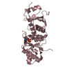
| Title | CRYSTAL STRUCTURE OF THE ARCHAEAL HOLLIDAY JUNCTION RESOLVASE HJC FROM PYROCOCCUS FURIOSUS FORM II | ||||||
 Components Components | HOLLIDAY JUNCTION RESOLVASE | ||||||
 Keywords Keywords | HYDROLASE / nuclease fold / HJC | ||||||
| Function / homology |  Function and homology information Function and homology informationcrossover junction endodeoxyribonuclease / Holliday junction resolvase complex / crossover junction DNA endonuclease activity / DNA recombination / DNA repair / magnesium ion binding / DNA binding Similarity search - Function | ||||||
| Biological species |   Pyrococcus furiosus (archaea) Pyrococcus furiosus (archaea) | ||||||
| Method |  X-RAY DIFFRACTION / SYNCHROTRON / MOLECULAR REPLACEMENT / Resolution: 2.16 Å X-RAY DIFFRACTION / SYNCHROTRON / MOLECULAR REPLACEMENT / Resolution: 2.16 Å | ||||||
 Authors Authors | Nishino, T. / Komori, K. / Ishino, Y. / Morikawa, K. | ||||||
 Citation Citation | Journal: J.Biol.Chem. / Year: 2001 Title: Dissection of the regional roles of the archaeal Holliday junction resolvase Hjc by structural and mutational analyses. Authors: Nishino, T. / Komori, K. / Ishino, Y. / Morikawa, K. | ||||||
| History |
|
- Structure visualization
| Structure viewer | Molecule: MolmilJmol/JSmol |
|---|
- Downloads & links
-Download
| PDBx/mmCIF format | 1ipi.cif.gz | 59.3 KB | Display | PDBx/mmCIF format |
|---|---|---|---|---|
| PDB format | pdb1ipi.ent.gz | 43.8 KB | Display | PDB format |
| PDBx/mmJSON format | 1ipi.json.gz | Tree view | PDBx/mmJSON format | |
| Others |  Other downloads Other downloads |
-Validation report
| Arichive directory | https://data.pdbj.org/pub/pdb/validation_reports/ip/1ipiftp://data.pdbj.org/pub/pdb/validation_reports/ip/1ipi | HTTPS FTP |
|---|
-Related structure data
| Related structure data |  1gefS S: Starting model for refinement |
|---|---|
| Similar structure data |


-Links
 PDBj
PDBj


- Assembly
| Deposited unit | 
| ||||||||
|---|---|---|---|---|---|---|---|---|---|
| 1 |
| ||||||||
| Unit cell |
| ||||||||
| Details | chain A and B form functional dimer |
-Components
| #1: Protein | Mass: 13792.379 Da / Num. of mol.: 2 Source method: isolated from a genetically manipulated source Source: (gene. exp.) Pyrococcus furiosus (archaea) / Gene: HJC / Plasmid: PET21 / Species (production host): Escherichia coli / Production host:  References: UniProt: Q9V301, UniProt: E7FHX4*PLUS, crossover junction endodeoxyribonuclease #2: Water | ChemComp-HOH / |  Mass: 18.015 Da / Num. of mol.: 157 / Source method: isolated from a natural source / Formula: H2O Mass: 18.015 Da / Num. of mol.: 157 / Source method: isolated from a natural source / Formula: H2O |
|---|
-Experimental details
-Experiment
| Experiment | Method: X-RAY DIFFRACTION / Number of used crystals: 1 |
|---|
- Sample preparation
| Crystal | Density Matthews: 2.53 Å3/Da / Density % sol: 51.41 % | ||||||||||||||||||||||||||||||||||||
|---|---|---|---|---|---|---|---|---|---|---|---|---|---|---|---|---|---|---|---|---|---|---|---|---|---|---|---|---|---|---|---|---|---|---|---|---|---|
| Crystal grow | Temperature: 297 K / Method: vapor diffusion / pH: 7 Details: 100mM Tris-HCl, 1mM EDTA, 32.5% PEG4000, 10% glycerol , pH 7.0, VAPOR DIFFUSION, temperature 297K | ||||||||||||||||||||||||||||||||||||
| Crystal grow | *PLUS Temperature: 20 ℃ / Method: batch method | ||||||||||||||||||||||||||||||||||||
| Components of the solutions | *PLUS
|
-Data collection
| Diffraction | Mean temperature: 100 K |
|---|---|
| Diffraction source | Source: SYNCHROTRON / Site: SPring-8  / Beamline: BL24XU / Wavelength: 0.834 Å / Beamline: BL24XU / Wavelength: 0.834 Å |
| Detector | Type: RIGAKU RAXIS / Detector: IMAGE PLATE / Date: Dec 11, 2000 |
| Radiation | Monochromator: DIAMOND / Protocol: SINGLE WAVELENGTH / Monochromatic (M) / Laue (L): M / Scattering type: x-ray |
| Radiation wavelength | Wavelength: 0.834 Å / Relative weight: 1 |
| Reflection | Resolution: 2.15→41.1 Å / Num. all: 94745 / Num. obs: 13043 / % possible obs: 88.9 % / Observed criterion σ(F): 1 / Observed criterion σ(I): 1 / Redundancy: 7.3 % / Biso Wilson estimate: 37 Å2 / Rmerge(I) obs: 0.069 / Net I/σ(I): 6.42 |
| Reflection shell | Resolution: 2.16→2.26 Å / Redundancy: 6 % / Rmerge(I) obs: 0.279 / Mean I/σ(I) obs: 2.7 / % possible all: 79.5 |
| Reflection | *PLUS Highest resolution: 2.16 Å / Lowest resolution: 41 Å / Rmerge(I) obs: 0.064 |
| Reflection shell | *PLUS Lowest resolution: 2.27 Å / % possible obs: 79.5 % |
- Processing
| Software |
| |||||||||||||||||||||||||
|---|---|---|---|---|---|---|---|---|---|---|---|---|---|---|---|---|---|---|---|---|---|---|---|---|---|---|
| Refinement | Method to determine structure: MOLECULAR REPLACEMENT Starting model: PDB ENTRY 1GEF functional dimer Resolution: 2.16→41 Å / Cross valid method: THROUGHOUT / Stereochemistry target values: Engh & Huber
| |||||||||||||||||||||||||
| Refine analyze |
| |||||||||||||||||||||||||
| Refinement step | Cycle: LAST / Resolution: 2.16→41 Å
| |||||||||||||||||||||||||
| Refine LS restraints |
| |||||||||||||||||||||||||
| LS refinement shell | Resolution: 2.16→2.24 Å
| |||||||||||||||||||||||||
| Software | *PLUS Name: CNS / Classification: refinement | |||||||||||||||||||||||||
| Refinement | *PLUS Lowest resolution: 41 Å / Num. reflection obs: 13043 / % reflection Rfree: 11.48 % / Rfactor obs: 0.214 | |||||||||||||||||||||||||
| Solvent computation | *PLUS | |||||||||||||||||||||||||
| Displacement parameters | *PLUS | |||||||||||||||||||||||||
| Refine LS restraints | *PLUS
| |||||||||||||||||||||||||
| LS refinement shell | *PLUS Lowest resolution: 2.27 Å / Rfactor Rfree: 0.307 / Rfactor Rwork: 0.233 / Rfactor obs: 0.233 |
