 Movie
Movie Controller


+ Open data

- Basic information
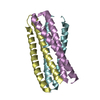
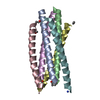
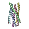
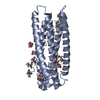
| Entry | Database: PDB / ID: 2siv | ||||||
|---|---|---|---|---|---|---|---|
| Title | SIV GP41 CORE STRUCTURE | ||||||
 Components Components | (SIV GP41 GLYCOPROTEIN) x 2 | ||||||
 Keywords Keywords | ENVELOPE GLYCOPROTEIN / RETROVIRUS / HIV / SIV / GP41 / COAT PROTEIN | ||||||
| Function / homology |  Function and homology information Function and homology informationmembrane fusion involved in viral entry into host cell / host cell endosome membrane / viral envelope / symbiont entry into host cell / virion attachment to host cell / host cell plasma membrane / virion membrane / structural molecule activity Similarity search - Function | ||||||
| Biological species |  Simian immunodeficiency virus Simian immunodeficiency virus | ||||||
| Method |  X-RAY DIFFRACTION / MOLECULAR REPLACEMENT / Resolution: 2.2 Å X-RAY DIFFRACTION / MOLECULAR REPLACEMENT / Resolution: 2.2 Å | ||||||
 Authors Authors | Malashkevich, V.N. / Chan, D.C. / Chutkowski, C.T. / Kim, P.S. | ||||||
 Citation Citation | Journal: Proc.Natl.Acad.Sci.USA / Year: 1998 Title: Crystal structure of the simian immunodeficiency virus (SIV) gp41 core: conserved helical interactions underlie the broad inhibitory activity of gp41 peptides. Authors: Malashkevich, V.N. / Chan, D.C. / Chutkowski, C.T. / Kim, P.S. #1: Journal: Cell(Cambridge,Mass.) / Year: 1997Title: Core Structure of Gp41 from the HIV Envelope Glycoprotein Authors: Chan, D.C. / Fass, D. / Berger, J.M. / Kim, P.S. | ||||||
| History |
|
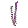
- Structure visualization
| Structure viewer | Molecule: MolmilJmol/JSmol |
|---|
- Downloads & links
-Download
| PDBx/mmCIF format | 2siv.cif.gz | 60.6 KB | Display | PDBx/mmCIF format |
|---|---|---|---|---|
| PDB format | pdb2siv.ent.gz | 45.4 KB | Display | PDB format |
| PDBx/mmJSON format | 2siv.json.gz | Tree view | PDBx/mmJSON format | |
| Others |  Other downloads Other downloads |
-Validation report
| Arichive directory | https://data.pdbj.org/pub/pdb/validation_reports/si/2sivftp://data.pdbj.org/pub/pdb/validation_reports/si/2siv | HTTPS FTP |
|---|
-Related structure data
| Related structure data | 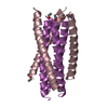 1aikS S: Starting model for refinement |
|---|---|
| Similar structure data |


-Links
 PDBj
PDBj


- Assembly
| Deposited unit | 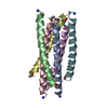
| ||||||||||||||||||||
|---|---|---|---|---|---|---|---|---|---|---|---|---|---|---|---|---|---|---|---|---|---|
| 1 |
| ||||||||||||||||||||
| Unit cell |
| ||||||||||||||||||||
| Noncrystallographic symmetry (NCS) | NCS oper:
|
-Components
| #1: Protein/peptide | Mass: 4218.902 Da / Num. of mol.: 3 / Fragment: PROTEASE-RESISTANT CORE Source method: isolated from a genetically manipulated source Details: N36 AND C34 ARE SYNTHETIC PEPTIDES / Source: (gene. exp.) Simian immunodeficiency virus / Genus: Lentivirus / Gene: GP41 / References: UniProt: Q87973, UniProt: Q8AKX0*PLUS#2: Protein/peptide | Mass: 4309.762 Da / Num. of mol.: 3 / Fragment: PROTEASE-RESISTANT CORE Source method: isolated from a genetically manipulated source Details: N36 AND C34 ARE SYNTHETIC PEPTIDES / Source: (gene. exp.) Simian immunodeficiency virus / Genus: Lentivirus / Strain: MAC239 / Cellular location: VIRAL MEMBRANE / Gene: GP41 / References: UniProt: Q87973, UniProt: Q52SW3*PLUS#3: Water | ChemComp-HOH / |  Mass: 18.015 Da / Num. of mol.: 184 / Source method: isolated from a natural source / Formula: H2O Mass: 18.015 Da / Num. of mol.: 184 / Source method: isolated from a natural source / Formula: H2OHas protein modification | Y | |
|---|
-Experimental details
-Experiment
| Experiment | Method: X-RAY DIFFRACTION / Number of used crystals: 1 |
|---|
- Sample preparation
| Crystal | Density Matthews: 2.2 Å3/Da / Density % sol: 44 % Description: CRYSTAL DEMONSTRATED HIGH MOSAICITY (1.2 DEGREES) AND ANISOTROPIC DIFFRACTION | |||||||||||||||||||||||||
|---|---|---|---|---|---|---|---|---|---|---|---|---|---|---|---|---|---|---|---|---|---|---|---|---|---|---|
| Crystal grow | pH: 6.5 Details: PROTEIN WAS CRYSTALLIZED FROM 18-19% PEG8000, 0.1 M SODIUM CACODILATE PH 6.5, 0.2 M MG-ACETATE | |||||||||||||||||||||||||
| Crystal | *PLUS | |||||||||||||||||||||||||
| Crystal grow | *PLUS Method: vapor diffusion, hanging drop | |||||||||||||||||||||||||
| Components of the solutions | *PLUS
|
-Data collection
| Diffraction | Mean temperature: 100 K |
|---|---|
| Diffraction source | Source: ROTATING ANODE / Type: RIGAKU RUH3R / Wavelength: 1.5418 |
| Detector | Type: RIGAKU RAXIS IV / Detector: IMAGE PLATE / Date: May 1, 1998 / Details: YALE MIRRORS |
| Radiation | Monochromator: NICKEL FILTER / Monochromatic (M) / Laue (L): M / Scattering type: x-ray |
| Radiation wavelength | Wavelength: 1.5418 Å / Relative weight: 1 |
| Reflection | Resolution: 2.2→25 Å / Num. obs: 11157 / % possible obs: 96.9 % / Observed criterion σ(I): 1 / Redundancy: 4.9 % / Biso Wilson estimate: 26.1 Å2 / Rmerge(I) obs: 0.058 / Rsym value: 0.058 / Net I/σ(I): 23.3 |
| Reflection shell | Resolution: 2.2→2.28 Å / Redundancy: 4.5 % / Rmerge(I) obs: 0.244 / Mean I/σ(I) obs: 5.5 / Rsym value: 0.244 / % possible all: 88.3 |
| Reflection | *PLUS Highest resolution: 2.2 Å / Num. measured all: 52353 |
- Processing
| Software |
| ||||||||||||||||||||||||||||||||||||||||||||||||||||||||||||||||||||||||||||||||
|---|---|---|---|---|---|---|---|---|---|---|---|---|---|---|---|---|---|---|---|---|---|---|---|---|---|---|---|---|---|---|---|---|---|---|---|---|---|---|---|---|---|---|---|---|---|---|---|---|---|---|---|---|---|---|---|---|---|---|---|---|---|---|---|---|---|---|---|---|---|---|---|---|---|---|---|---|---|---|---|---|---|
| Refinement | Method to determine structure: MOLECULAR REPLACEMENT Starting model: PDB ENTRY 1AIK Resolution: 2.2→20 Å / Rfactor Rfree error: 0.009 / Data cutoff high absF: 10000000 / Data cutoff low absF: 0.001 / Isotropic thermal model: RESTRAINED / Cross valid method: THROUGHOUT / σ(F): 2 / Details: BULK SOLVENT MODEL USED
| ||||||||||||||||||||||||||||||||||||||||||||||||||||||||||||||||||||||||||||||||
| Displacement parameters | Biso mean: 35.4 Å2
| ||||||||||||||||||||||||||||||||||||||||||||||||||||||||||||||||||||||||||||||||
| Refine analyze |
| ||||||||||||||||||||||||||||||||||||||||||||||||||||||||||||||||||||||||||||||||
| Refinement step | Cycle: LAST / Resolution: 2.2→20 Å
| ||||||||||||||||||||||||||||||||||||||||||||||||||||||||||||||||||||||||||||||||
| Refine LS restraints |
| ||||||||||||||||||||||||||||||||||||||||||||||||||||||||||||||||||||||||||||||||
| LS refinement shell | Resolution: 2.2→2.34 Å / Rfactor Rfree error: 0.033 / Total num. of bins used: 6
| ||||||||||||||||||||||||||||||||||||||||||||||||||||||||||||||||||||||||||||||||
| Xplor file |
| ||||||||||||||||||||||||||||||||||||||||||||||||||||||||||||||||||||||||||||||||
| Software | *PLUS Name: X-PLOR / Version: 3.851 / Classification: refinement | ||||||||||||||||||||||||||||||||||||||||||||||||||||||||||||||||||||||||||||||||
| Refine LS restraints | *PLUS
| ||||||||||||||||||||||||||||||||||||||||||||||||||||||||||||||||||||||||||||||||
| LS refinement shell | *PLUS Rfactor Rwork: 0.33 |
