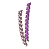
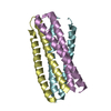
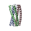
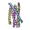
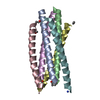
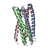
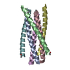
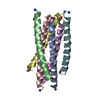
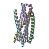
VIRAL PROTEIN / HIV / GP41 / ENVELOPE GLYCOPROTEIN / RETROVIRUS
Function / homology
Function and homology information
Synthesis and processing of ENV and VPU / symbiont-mediated evasion of host immune response / positive regulation of establishment of T cell polarity / Alpha-defensins / Dectin-2 family / Binding and entry of HIV virion / positive regulation of plasma membrane raft polarization / positive regulation of receptor clustering / host cell endosome membrane / actin filament organization ...Synthesis and processing of ENV and VPU / symbiont-mediated evasion of host immune response / positive regulation of establishment of T cell polarity / Alpha-defensins / Dectin-2 family / Binding and entry of HIV virion / positive regulation of plasma membrane raft polarization / positive regulation of receptor clustering / host cell endosome membrane / actin filament organization / Assembly Of The HIV Virion / Budding and maturation of HIV virion / clathrin-dependent endocytosis of virus by host cell / viral protein processing / receptor ligand activity / fusion of virus membrane with host plasma membrane / fusion of virus membrane with host endosome membrane / viral envelope / symbiont entry into host cell / virion attachment to host cell / host cell plasma membrane / virion membrane / structural molecule activity / membrane Similarity search - Function
Mass: 18.015 Da / Num. of mol.: 43 / Source method: isolated from a natural source / Formula: H2O
Has protein modification
Y
-
Experimental details
-
Experiment
Experiment
Method: X-RAY DIFFRACTION / Number of used crystals: 1
-
Sample preparation
Crystal
Density Matthews: 2.32 Å3/Da / Density % sol: 46 % Description: DATA AT NSLS USED MAD METHODS. DATA COLLECTED ON AN OSMIUM-SOAK CRYSTAL AT WAVELENGTHS 1.1398, 1.1396, 1.1344, AND 1.1406 ANGSTROMS.
Crystal grow
Method: vapor diffusion, sitting drop / pH: 6 Details: A 10 MG/ML STOCK WAS DILUTED 1:1 IN A SITTING DROP WITH 80 MM NH4CL, 20% PEG200, AND 50% ISOPROPANOL, AND THEN ALLOWED TO EQUILIBRATE AGAINST 80 MM NH4CL, 20% PEG200, AND 30% ISOPROPANOL., ...Details: A 10 MG/ML STOCK WAS DILUTED 1:1 IN A SITTING DROP WITH 80 MM NH4CL, 20% PEG200, AND 50% ISOPROPANOL, AND THEN ALLOWED TO EQUILIBRATE AGAINST 80 MM NH4CL, 20% PEG200, AND 30% ISOPROPANOL., pH 6.0, vapor diffusion - sitting drop
In the structure databanks used in Yorodumi, some data are registered as the other names, "COVID-19 virus" and "2019-nCoV". Here are the details of the virus and the list of structure data.
Jan 31, 2019. EMDB accession codes are about to change! (news from PDBe EMDB page)
EMDB accession codes are about to change! (news from PDBe EMDB page)
The allocation of 4 digits for EMDB accession codes will soon come to an end. Whilst these codes will remain in use, new EMDB accession codes will include an additional digit and will expand incrementally as the available range of codes is exhausted. The current 4-digit format prefixed with “EMD-” (i.e. EMD-XXXX) will advance to a 5-digit format (i.e. EMD-XXXXX), and so on. It is currently estimated that the 4-digit codes will be depleted around Spring 2019, at which point the 5-digit format will come into force.
The EM Navigator/Yorodumi systems omit the EMD- prefix.
Related info.:Q: What is EMD? / ID/Accession-code notation in Yorodumi/EM Navigator
Yorodumi is a browser for structure data from EMDB, PDB, SASBDB, etc.
This page is also the successor to EM Navigator detail page, and also detail information page/front-end page for Omokage search.
The word "yorodu" (or yorozu) is an old Japanese word meaning "ten thousand". "mi" (miru) is to see.
Related info.:EMDB / PDB / SASBDB / Comparison of 3 databanks / Yorodumi Search / Aug 31, 2016. New EM Navigator & Yorodumi / Yorodumi Papers / Jmol/JSmol / Function and homology information / Changes in new EM Navigator and Yorodumi
 Movie
Movie Controller


 Open data
 Basic information
 Components
 Keywords
 Function and homology information
 HIV-1 M:B_HXB2R (virus)
 X-RAY DIFFRACTION /
  Authors
 Citation
 Structure visualization
 Downloads & links
 Other downloads
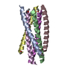
 PDBj
PDBj


 Assembly
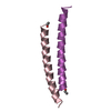


 Mass: 18.015 Da / Num. of mol.: 43 / Source method: isolated from a natural source / Formula: H2O
 Sample preparation
 Processing