 Movie
Movie Controller

[English] 日本語
 Yorodumi
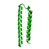
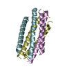
- PDB-2ot5: Crystal structure of the HIV gp41 core with the enfuvirtide resis... -
+ Open data

- Basic information
| Entry | Database: PDB / ID: 2ot5 | ||||||
|---|---|---|---|---|---|---|---|
| Title | Crystal structure of the HIV gp41 core with the enfuvirtide resistance mutation N43D | ||||||
 Components Components | HIV-1 gp41 glycoprotein | ||||||
 Keywords Keywords | VIRAL PROTEIN / gp41 / coiled-coil | ||||||
| Function / homology |  Function and homology information Function and homology informationmembrane fusion involved in viral entry into host cell / viral envelope / symbiont entry into host cell / virion attachment to host cell / host cell plasma membrane / virion membrane / structural molecule activity Similarity search - Function | ||||||
| Biological species |   human immunodeficiency virus type 1 human immunodeficiency virus type 1 | ||||||
| Method |  X-RAY DIFFRACTION / MOLECULAR REPLACEMENT / Resolution: 1.8 Å X-RAY DIFFRACTION / MOLECULAR REPLACEMENT / Resolution: 1.8 Å | ||||||
 Authors Authors | Bai, X. / Seedorff, J.E. / Green, J. / Dwyer, J.J. | ||||||
 Citation Citation | Journal: Biochemistry / Year: 2008 Title: Impact of the enfuvirtide resistance mutation N43D and the associated baseline polymorphism E137K on peptide sensitivity and six-helix bundle structure. Authors: Bai, X. / Wilson, K.L. / Seedorff, J.E. / Ahrens, D. / Green, J. / Davison, D.K. / Jin, L. / Stanfield-Oakley, S.A. / Mosier, S.M. / Melby, T.E. / Cammack, N. / Wang, Z. / Greenberg, M.L. / Dwyer, J.J. | ||||||
| History |
|
- Structure visualization
| Structure viewer | Molecule: MolmilJmol/JSmol |
|---|
- Downloads & links
-Download
| PDBx/mmCIF format | 2ot5.cif.gz | 25.1 KB | Display | PDBx/mmCIF format |
|---|---|---|---|---|
| PDB format | pdb2ot5.ent.gz | 16.2 KB | Display | PDB format |
| PDBx/mmJSON format | 2ot5.json.gz | Tree view | PDBx/mmJSON format | |
| Others |  Other downloads Other downloads |
-Validation report
| Arichive directory | https://data.pdbj.org/pub/pdb/validation_reports/ot/2ot5ftp://data.pdbj.org/pub/pdb/validation_reports/ot/2ot5 | HTTPS FTP |
|---|
-Related structure data
| Related structure data | 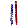 3cp1C 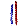 3cyoC  1stzS S: Starting model for refinement C: citing same article ( |
|---|---|
| Similar structure data |


-Links
 PDBj
PDBj


- Assembly
| Deposited unit | 
| ||||||||
|---|---|---|---|---|---|---|---|---|---|
| 1 | 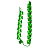
| ||||||||
| Unit cell |
|
-Components
| #1: Protein | Mass: 7880.738 Da / Num. of mol.: 1 / Fragment: Subdomain N34(L6)C28 / Mutation: N43D Source method: isolated from a genetically manipulated source Source: (gene. exp.) human immunodeficiency virus type 1 / Strain: hxb2 / Plasmid: pET4a / Production host:  |
|---|---|
| #2: Water | ChemComp-HOH /  Mass: 18.015 Da / Num. of mol.: 25 / Source method: isolated from a natural source / Formula: H2O Mass: 18.015 Da / Num. of mol.: 25 / Source method: isolated from a natural source / Formula: H2O |
-Experimental details
-Experiment
| Experiment | Method: X-RAY DIFFRACTION / Number of used crystals: 1 |
|---|
- Sample preparation
| Crystal | Density Matthews: 1.9 Å3/Da / Density % sol: 35.15 % |
|---|---|
| Crystal grow | Temperature: 298 K / Method: vapor diffusion, hanging drop / pH: 4.3 Details: 0.05M sodium acetate, 0.2M ammonium sulfate, 23% PEG 4000, 10% glycerol, pH 4.3, VAPOR DIFFUSION, HANGING DROP, temperature 298K |
-Data collection
| Diffraction | Mean temperature: 298 K |
|---|---|
| Diffraction source | Source: ROTATING ANODE / Type: RIGAKU RU200 / Wavelength: 1.5418 |
| Detector | Type: RIGAKU RAXIS IV / Detector: IMAGE PLATE / Date: Jul 5, 2005 |
| Radiation | Monochromator: graphite / Protocol: SINGLE WAVELENGTH / Monochromatic (M) / Laue (L): M / Scattering type: x-ray |
| Radiation wavelength | Wavelength: 1.5418 Å / Relative weight: 1 |
| Reflection | Resolution: 1.56→17.76 Å / Num. all: 8182 / Num. obs: 6929 / % possible obs: 84.7 % / Observed criterion σ(F): 2 / Observed criterion σ(I): 2 / Redundancy: 4.95 % / Rmerge(I) obs: 0.035 / Χ2: 0.94 / Net I/σ(I): 30.9 / Scaling rejects: 261 |
| Reflection shell | Resolution: 1.56→1.62 Å / Redundancy: 1.52 % / Rmerge(I) obs: 0.502 / Mean I/σ(I) obs: 2 / Num. measured all: 219 / Num. unique all: 144 / Χ2: 0.47 / % possible all: 17.2 |
- Processing
| Software |
| ||||||||||||||||||||||||||||
|---|---|---|---|---|---|---|---|---|---|---|---|---|---|---|---|---|---|---|---|---|---|---|---|---|---|---|---|---|---|
| Refinement | Method to determine structure: MOLECULAR REPLACEMENT Starting model: pdb entry 1STZ Resolution: 1.8→17.76 Å / Isotropic thermal model: anisotropic / σ(F): 0 / Stereochemistry target values: Engh & Huber
| ||||||||||||||||||||||||||||
| Solvent computation | Bsol: 53.988 Å2 | ||||||||||||||||||||||||||||
| Displacement parameters | Biso mean: 34.141 Å2
| ||||||||||||||||||||||||||||
| Refinement step | Cycle: LAST / Resolution: 1.8→17.76 Å
| ||||||||||||||||||||||||||||
| Refine LS restraints |
| ||||||||||||||||||||||||||||
| LS refinement shell | Resolution: 2→2.09 Å / Rfactor Rfree error: 0.024
| ||||||||||||||||||||||||||||
| Xplor file |
|
