 Movie
Movie Controller


+ Open data

- Basic information
| Entry | Database: PDB / ID: 1szt | ||||||
|---|---|---|---|---|---|---|---|
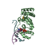
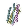
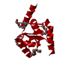
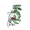
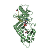
| Title | ATOMIC STRUCTURE OF A THERMOSTABLE SUBDOMAIN OF HIV-1 GP41 | ||||||
 Components Components | HIV-1 ENVELOPE GLYCOPROTEIN GP41 | ||||||
 Keywords Keywords | VIRAL PROTEIN / COAT PROTEIN / HIV-1 ENVELOPE GLYCOPROTEIN | ||||||
| Function / homology |  Function and homology information Function and homology informationDectin-2 family / symbiont-mediated perturbation of host defense response / positive regulation of plasma membrane raft polarization / positive regulation of receptor clustering / host cell endosome membrane / clathrin-dependent endocytosis of virus by host cell / viral protein processing / fusion of virus membrane with host plasma membrane / fusion of virus membrane with host endosome membrane / viral envelope ...Dectin-2 family / symbiont-mediated perturbation of host defense response / positive regulation of plasma membrane raft polarization / positive regulation of receptor clustering / host cell endosome membrane / clathrin-dependent endocytosis of virus by host cell / viral protein processing / fusion of virus membrane with host plasma membrane / fusion of virus membrane with host endosome membrane / viral envelope / symbiont entry into host cell / virion attachment to host cell / host cell plasma membrane / virion membrane / structural molecule activity / membrane Similarity search - Function | ||||||
| Biological species |   Human immunodeficiency virus 1 Human immunodeficiency virus 1 | ||||||
| Method |  X-RAY DIFFRACTION / SIRAS / Resolution: 2.4 Å X-RAY DIFFRACTION / SIRAS / Resolution: 2.4 Å | ||||||
 Authors Authors | Tan, K. / Lu, M. / Wang, J.-H. | ||||||
 Citation Citation | Journal: Proc.Natl.Acad.Sci.USA / Year: 1997 Title: Atomic structure of a thermostable subdomain of HIV-1 gp41. Authors: Tan, K. / Liu, J. / Wang, J. / Shen, S. / Lu, M. #1: Journal: Cell(Cambridge,Mass.) / Year: 1997Title: Core Structure of Gp41 from the HIV Envelope Glycoprotein Authors: Chan, D.C. / Fass, D. / Berger, J.M. / Kim, P.S. #2: Journal: Nature / Year: 1997Title: Atomic Structure of the Ectodomain from HIV-1 Gp41 Authors: Weissenhorn, W. / Dessen, A. / Harrison, S.C. / Skehel, J.J. / Wiley, D.C. #3: Journal: Nat.Struct.Biol. / Year: 1995Title: A Trimeric Structural Domain of the HIV-1 Transmembrane Glycoprotein Authors: Lu, M. / Blacklow, S.C. / Kim, P.S. | ||||||
| History |
|
- Structure visualization
| Structure viewer | Molecule: MolmilJmol/JSmol |
|---|
- Downloads & links
-Download
| PDBx/mmCIF format | 1szt.cif.gz | 25.4 KB | Display | PDBx/mmCIF format |
|---|---|---|---|---|
| PDB format | pdb1szt.ent.gz | 17 KB | Display | PDB format |
| PDBx/mmJSON format | 1szt.json.gz | Tree view | PDBx/mmJSON format | |
| Others |  Other downloads Other downloads |
-Validation report
| Arichive directory | https://data.pdbj.org/pub/pdb/validation_reports/sz/1sztftp://data.pdbj.org/pub/pdb/validation_reports/sz/1szt | HTTPS FTP |
|---|
-Related structure data
| Similar structure data |
|---|


-Links
 PDBj
PDBj


- Assembly
| Deposited unit | 
| ||||||||
|---|---|---|---|---|---|---|---|---|---|
| 1 | 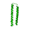
| ||||||||
| Unit cell |
|
-Components
| #1: Protein | Mass: 7879.753 Da / Num. of mol.: 1 Fragment: RESIDUES 546 - 579 AND 628 - 655 CONNECTED BY A SIX-RESIDUE LINKER (SER-GLY-GLY-ARG-GLY-GLY) Source method: isolated from a genetically manipulated source Details: CORE OF TRANSMEMBRANE SUBUNIT (GP41) / Source: (gene. exp.) Human immunodeficiency virus 1 / Genus: LentivirusDescription: RECOMBINANT GP41 WITH LINKER (SER-GLY-GLY-ARG-GLY-GLY) BETWEEN TWO FRAGMENTS Cell line: BL21 / Gene: GP41 / Plasmid: PN41/C34-L6 / Gene (production host): GP41 / Production host:  |
|---|---|
| #2: Water | ChemComp-HOH /  Mass: 18.015 Da / Num. of mol.: 42 / Source method: isolated from a natural source / Formula: H2O Mass: 18.015 Da / Num. of mol.: 42 / Source method: isolated from a natural source / Formula: H2O |
| Compound details | IN THE STRUCTURE, SEQUENCE 1 - 34 IS FROM GP41 RESIDUES 546 - 579 (IN GP160 NUMBERING SYSTEM), 31 - ...IN THE STRUCTURE, SEQUENCE 1 - 34 IS FROM GP41 RESIDUES 546 - 579 (IN GP160 NUMBERING SYSTEM), 31 - 40 IS AN ARTIFICIAL |
-Experimental details
-Experiment
| Experiment | Method: X-RAY DIFFRACTION / Number of used crystals: 1 |
|---|
- Sample preparation
| Crystal | Density Matthews: 2.08 Å3/Da / Density % sol: 40 % | ||||||||||||||||||||||||||||||||||||
|---|---|---|---|---|---|---|---|---|---|---|---|---|---|---|---|---|---|---|---|---|---|---|---|---|---|---|---|---|---|---|---|---|---|---|---|---|---|
| Crystal grow | *PLUS Temperature: 20 ℃ / pH: 5 / Method: vapor diffusion, hanging dropDetails: drop solution was mixed with an equal volume of reservoir solution | ||||||||||||||||||||||||||||||||||||
| Components of the solutions | *PLUS
|
-Data collection
| Diffraction | Mean temperature: 293 K |
|---|---|
| Diffraction source | Wavelength: 1.5418 |
| Detector | Type: MARRESEARCH / Detector: IMAGE PLATE / Date: Feb 26, 1997 |
| Radiation | Monochromatic (M) / Laue (L): M / Scattering type: x-ray |
| Radiation wavelength | Wavelength: 1.5418 Å / Relative weight: 1 |
| Reflection | Resolution: 2.4→15 Å / Num. obs: 2481 / % possible obs: 99.6 % / Observed criterion σ(I): -3 / Redundancy: 8.9 % / Rmerge(I) obs: 0.057 / Net I/σ(I): 27.61 |
| Reflection shell | Resolution: 2.4→2.49 Å / Redundancy: 6.2 % / Rmerge(I) obs: 0.211 / Mean I/σ(I) obs: 8.98 / % possible all: 99.6 |
| Reflection shell | *PLUS % possible obs: 99.6 % |
- Processing
| Software |
| ||||||||||||||||||||||||||||||||||||||||||||||||||||||||||||
|---|---|---|---|---|---|---|---|---|---|---|---|---|---|---|---|---|---|---|---|---|---|---|---|---|---|---|---|---|---|---|---|---|---|---|---|---|---|---|---|---|---|---|---|---|---|---|---|---|---|---|---|---|---|---|---|---|---|---|---|---|---|
| Refinement | Method to determine structure: SIRAS / Resolution: 2.4→15 Å / σ(F): 3
| ||||||||||||||||||||||||||||||||||||||||||||||||||||||||||||
| Displacement parameters | Biso mean: 61.06 Å2 | ||||||||||||||||||||||||||||||||||||||||||||||||||||||||||||
| Refine analyze | Luzzati coordinate error obs: 0.34 Å | ||||||||||||||||||||||||||||||||||||||||||||||||||||||||||||
| Refinement step | Cycle: LAST / Resolution: 2.4→15 Å
| ||||||||||||||||||||||||||||||||||||||||||||||||||||||||||||
| Refine LS restraints |
| ||||||||||||||||||||||||||||||||||||||||||||||||||||||||||||
| Software | *PLUS Name: X-PLOR / Classification: refinement | ||||||||||||||||||||||||||||||||||||||||||||||||||||||||||||
| Refinement | *PLUS | ||||||||||||||||||||||||||||||||||||||||||||||||||||||||||||
| Solvent computation | *PLUS | ||||||||||||||||||||||||||||||||||||||||||||||||||||||||||||
| Displacement parameters | *PLUS |
