Mass: 18.015 Da / Num. of mol.: 388 / Source method: isolated from a natural source / Formula: H2O
Sequence details
AUTHORS STATE THAT RESIDUE 269 IS LEU ACCORDING TO THE DENSITY MAPS.
-
Experimental details
-
Experiment
Experiment
Method: X-RAY DIFFRACTION / Number of used crystals: 1
-
Sample preparation
Crystal
Density Matthews: 2.23 Å3/Da / Density % sol: 44.9 %
Crystal grow
Temperature: 298 K / Method: vapor diffusion, hanging drop Details: protein: 9.1 mg/mL in 20 mM Tris-HCl, pH 8.0, 0.1M KCl, 10 mM pyruvate. precipitant: 24%(w/v) PEG6000, 50 mM NaH2PO4. 10 uL droplets (5uL protein + 5 uL precipitant) suspended over 1mL of ...Details: protein: 9.1 mg/mL in 20 mM Tris-HCl, pH 8.0, 0.1M KCl, 10 mM pyruvate. precipitant: 24%(w/v) PEG6000, 50 mM NaH2PO4. 10 uL droplets (5uL protein + 5 uL precipitant) suspended over 1mL of precipitant solution, VAPOR DIFFUSION, HANGING DROP, temperature 298K
Starting model: PDB entry 1DHP was used to get an initial MR structure solution. A better MR starting solution resulted later when PDB entry 1XXX was made available for use.
Resolution: 2.2→20 Å / σ(F): 0 / Stereochemistry target values: Engh & Huber Details: The active site lysine residues (176a, 176b) form a Schiff base complex with pyruvate. Proline residues 282a, 282b are cis-prolines.
Rfactor
Num. reflection
% reflection
Selection details
Rfree
0.259
2680
9.4 %
10% of data
Rwork
0.212
-
-
-
obs
0.228
26969
95.7 %
-
all
-
27767
-
-
Displacement parameters
Biso mean: 13.105 Å2
Refine analyze
Luzzati coordinate error obs: 0.313 Å / Luzzati d res low obs: 5 Å
Refinement step
Cycle: LAST / Resolution: 2.2→20 Å
Protein
Nucleic acid
Ligand
Solvent
Total
Num. atoms
4418
0
0
388
4806
Refine LS restraints
Refine-ID
Type
Dev ideal
Dev ideal target
X-RAY DIFFRACTION
x_bond_d
0.009
1.5
X-RAY DIFFRACTION
x_angle_deg
1.516
2
LS refinement shell
Resolution (Å)
Rfactor Rfree
Num. reflection Rfree
Rfactor Rwork
Refine-ID
Num. reflection obs
2.2-2.34
0.2599
418
0.211
X-RAY DIFFRACTION
4120
2.34-2.52
0.2375
418
0.1824
X-RAY DIFFRACTION
4375
2.52-2.77
0.2596
462
0.2066
X-RAY DIFFRACTION
4482
2.77-3.17
0.2615
457
0.2019
X-RAY DIFFRACTION
4571
3.17-3.99
0.2345
460
0.1889
X-RAY DIFFRACTION
4695
3.99-20
0.284
465
0.2531
X-RAY DIFFRACTION
4726
Xplor file
Refine-ID
Serial no
Param file
X-RAY DIFFRACTION
1
protein_rep_mcl7.param
X-RAY DIFFRACTION
2
dna-rna_rep.param
X-RAY DIFFRACTION
3
water_rep.param
X-RAY DIFFRACTION
4
ion.param
+
About Yorodumi
-
News
-
Feb 9, 2022. New format data for meta-information of EMDB entries
New format data for meta-information of EMDB entries
Version 3 of the EMDB header file is now the official format.
The previous official version 1.9 will be removed from the archive.
In the structure databanks used in Yorodumi, some data are registered as the other names, "COVID-19 virus" and "2019-nCoV". Here are the details of the virus and the list of structure data.
Jan 31, 2019. EMDB accession codes are about to change! (news from PDBe EMDB page)
EMDB accession codes are about to change! (news from PDBe EMDB page)
The allocation of 4 digits for EMDB accession codes will soon come to an end. Whilst these codes will remain in use, new EMDB accession codes will include an additional digit and will expand incrementally as the available range of codes is exhausted. The current 4-digit format prefixed with “EMD-” (i.e. EMD-XXXX) will advance to a 5-digit format (i.e. EMD-XXXXX), and so on. It is currently estimated that the 4-digit codes will be depleted around Spring 2019, at which point the 5-digit format will come into force.
The EM Navigator/Yorodumi systems omit the EMD- prefix.
Related info.:Q: What is EMD? / ID/Accession-code notation in Yorodumi/EM Navigator
Yorodumi is a browser for structure data from EMDB, PDB, SASBDB, etc.
This page is also the successor to EM Navigator detail page, and also detail information page/front-end page for Omokage search.
The word "yorodu" (or yorozu) is an old Japanese word meaning "ten thousand". "mi" (miru) is to see.
Related info.:EMDB / PDB / SASBDB / Comparison of 3 databanks / Yorodumi Search / Aug 31, 2016. New EM Navigator & Yorodumi / Yorodumi Papers / Jmol/JSmol / Function and homology information / Changes in new EM Navigator and Yorodumi
 Movie
Movie Controller

 Yorodumi
 Open data
 Basic information
 Components
 Keywords
 Function and homology information
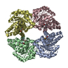
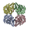
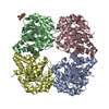
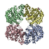
 Corynebacterium glutamicum (bacteria)
 X-RAY DIFFRACTION /
  Authors
 Citation
 Structure visualization
 Downloads & links
 Other downloads


 PDBj
PDBj Assembly
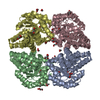
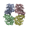
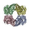
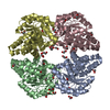
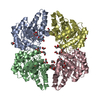
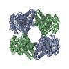
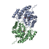
 Mass: 18.015 Da / Num. of mol.: 388 / Source method: isolated from a natural source / Formula: H2O
 Sample preparation
 / Beamline: 22-ID / Wavelength: 1 Å
 Processing