 Movie
Movie Controller

[English] 日本語
 Yorodumi
- PDB-4bwl: Structure of the Y137A mutant of E. coli N-acetylneuraminic acid ... -
+ Open data

- Basic information
| Entry | Database: PDB / ID: 4bwl | ||||||
|---|---|---|---|---|---|---|---|
| Title | Structure of the Y137A mutant of E. coli N-acetylneuraminic acid lyase in complex with pyruvate, N-acetyl-D-mannosamine and N- acetylneuraminic acid | ||||||
 Components Components | (N-ACETYLNEURAMINATE ...) x 2 | ||||||
 Keywords Keywords | LYASE / DIRECTED EVOLUTION / SUBSTRATE SPECIFICITY / PROTEIN ENGINEERING | ||||||
| Function / homology |  Function and homology information Function and homology informationN-acetylneuraminate lyase / N-acetylneuraminate lyase activity / N-acetylneuraminate catabolic process / single-species biofilm formation / carbohydrate metabolic process / identical protein binding / cytosol Similarity search - Function | ||||||
| Biological species |  | ||||||
| Method |  X-RAY DIFFRACTION / SYNCHROTRON / MOLECULAR REPLACEMENT / Resolution: 2 Å X-RAY DIFFRACTION / SYNCHROTRON / MOLECULAR REPLACEMENT / Resolution: 2 Å | ||||||
 Authors Authors | Campeotto, I. / Phillips, S.E.V. / Pearson, A.R. / Nelson, A. / Berry, A. | ||||||
 Citation Citation | Journal: Acs Chem.Biol. / Year: 2014 Title: The Reaction Mechanism of N-Acetylneuraminic Acid Lyase Revealed by a Combination of Crystallography, Qm/Mm Simulation and Mutagenesis. Authors: Daniels, A.D. / Campeotto, I. / Van Der Kamp, M.W. / Bolt, A.H. / Trinh, C.H. / Phillips, S.E.V. / Pearson, A.R. / Nelson, A. / Mulholland, A.J. / Berry, A. | ||||||
| History |
| ||||||
| Remark 700 | SHEET DETERMINATION METHOD: DSSP THE SHEETS PRESENTED AS "AA" IN EACH CHAIN ON SHEET RECORDS BELOW ... SHEET DETERMINATION METHOD: DSSP THE SHEETS PRESENTED AS "AA" IN EACH CHAIN ON SHEET RECORDS BELOW IS ACTUALLY AN 8-STRANDED BARREL THIS IS REPRESENTED BY A 9-STRANDED SHEET IN WHICH THE FIRST AND LAST STRANDS ARE IDENTICAL. THE SHEETS PRESENTED AS "BA" IN EACH CHAIN ON SHEET RECORDS BELOW IS ACTUALLY AN 8-STRANDED BARREL THIS IS REPRESENTED BY A 9-STRANDED SHEET IN WHICH THE FIRST AND LAST STRANDS ARE IDENTICAL. THE SHEETS PRESENTED AS "CA" IN EACH CHAIN ON SHEET RECORDS BELOW IS ACTUALLY AN 8-STRANDED BARREL THIS IS REPRESENTED BY A 9-STRANDED SHEET IN WHICH THE FIRST AND LAST STRANDS ARE IDENTICAL. THE SHEETS PRESENTED AS "DA" IN EACH CHAIN ON SHEET RECORDS BELOW IS ACTUALLY AN 8-STRANDED BARREL THIS IS REPRESENTED BY A 9-STRANDED SHEET IN WHICH THE FIRST AND LAST STRANDS ARE IDENTICAL. |
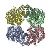
- Structure visualization
| Structure viewer | Molecule: MolmilJmol/JSmol |
|---|
- Downloads & links
-Download
| PDBx/mmCIF format | 4bwl.cif.gz | 235.6 KB | Display | PDBx/mmCIF format |
|---|---|---|---|---|
| PDB format | pdb4bwl.ent.gz | 188.6 KB | Display | PDB format |
| PDBx/mmJSON format | 4bwl.json.gz | Tree view | PDBx/mmJSON format | |
| Others |  Other downloads Other downloads |
-Validation report
| Arichive directory | https://data.pdbj.org/pub/pdb/validation_reports/bw/4bwlftp://data.pdbj.org/pub/pdb/validation_reports/bw/4bwl | HTTPS FTP |
|---|
-Related structure data
| Related structure data |  2wnnS S: Starting model for refinement |
|---|---|
| Similar structure data |


-Links
 PDBj
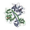
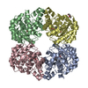
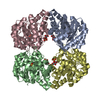
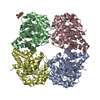
PDBj- Assembly
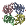
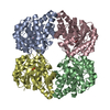
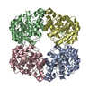
| Deposited unit | 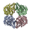
| ||||||||
|---|---|---|---|---|---|---|---|---|---|
| 1 |
| ||||||||
| Unit cell |
|
-Components
-N-ACETYLNEURAMINATE ... , 2 types, 4 molecules ABDC
| #1: Protein | Mass: 33492.273 Da / Num. of mol.: 3 / Mutation: YES Source method: isolated from a genetically manipulated source Details: SCHIFF BASE BETWEEN LYS165 AND N-ACETYLNEURAMINIC ACID Source: (gene. exp.) #2: Protein | | Mass: 33561.312 Da / Num. of mol.: 1 / Mutation: YES Source method: isolated from a genetically manipulated source Details: SCHIFF BASE BETWEEN LYS165 AND PYRUVATE / Source: (gene. exp.) |
|---|
-Sugars , 1 types, 3 molecules 
| #3: Sugar |  Type: D-saccharide / Mass: 309.270 Da / Num. of mol.: 3 Type: D-saccharide / Mass: 309.270 Da / Num. of mol.: 3Source method: isolated from a genetically manipulated source Formula: C11H19NO9 |
|---|
-Non-polymers , 3 types, 105 molecules 


| #4: Chemical |  Mass: 238.278 Da / Num. of mol.: 2 / Source method: obtained synthetically / Formula: C10H22O6 / Comment: precipitant*YM Mass: 238.278 Da / Num. of mol.: 2 / Source method: obtained synthetically / Formula: C10H22O6 / Comment: precipitant*YM#5: Chemical | ChemComp-MN9 / |  Mass: 221.208 Da / Num. of mol.: 1 / Source method: obtained synthetically / Formula: C8H15NO6 Mass: 221.208 Da / Num. of mol.: 1 / Source method: obtained synthetically / Formula: C8H15NO6#6: Water | ChemComp-HOH / | Mass: 18.015 Da / Num. of mol.: 102 / Source method: isolated from a natural source / Formula: H2O |
|---|
-Experimental details
-Experiment
| Experiment | Method: X-RAY DIFFRACTION / Number of used crystals: 1 |
|---|
- Sample preparation
| Crystal | Density Matthews: 2.42 Å3/Da / Density % sol: 49.29 % / Description: NONE |
|---|---|
| Crystal grow | pH: 8.2 / Details: 100MM TRIS-HCL PH 8.2, 200MM NACL, 18% PEG3350 |
-Data collection
| Diffraction | Mean temperature: 100 K | |||||||||||||||
|---|---|---|---|---|---|---|---|---|---|---|---|---|---|---|---|---|
| Diffraction source | Source: SYNCHROTRON / Site: Diamond  / Beamline: I02 / Wavelength: 0.97 / Beamline: I02 / Wavelength: 0.97 | |||||||||||||||
| Detector | Type: ADSC CCD / Detector: CCD / Date: Jul 8, 2009 | |||||||||||||||
| Radiation | Protocol: SINGLE WAVELENGTH / Monochromatic (M) / Laue (L): M / Scattering type: x-ray | |||||||||||||||
| Radiation wavelength | Wavelength: 0.97 Å / Relative weight: 1 | |||||||||||||||
| Reflection twin |
| |||||||||||||||
| Reflection | Resolution: 2→49.5 Å / Num. obs: 82202 / % possible obs: 98.7 % / Observed criterion σ(I): 2 / Redundancy: 3.6 % / Biso Wilson estimate: 21.4 Å2 / Rmerge(I) obs: 0.09 / Net I/σ(I): 9.6 | |||||||||||||||
| Reflection shell | Resolution: 2→2.11 Å / Redundancy: 3.5 % / Rmerge(I) obs: 0.41 / Mean I/σ(I) obs: 2.8 / % possible all: 95.7 |
- Processing
| Software |
| ||||||||||||||||||||||||||||||||||||||||||||||||||||||||||||||||||||||||||||||||||||||||||||||||||||||||||||||||||||||||||||||||||||||||||||||||||||||||||||||||||||||||||||||||||||||
|---|---|---|---|---|---|---|---|---|---|---|---|---|---|---|---|---|---|---|---|---|---|---|---|---|---|---|---|---|---|---|---|---|---|---|---|---|---|---|---|---|---|---|---|---|---|---|---|---|---|---|---|---|---|---|---|---|---|---|---|---|---|---|---|---|---|---|---|---|---|---|---|---|---|---|---|---|---|---|---|---|---|---|---|---|---|---|---|---|---|---|---|---|---|---|---|---|---|---|---|---|---|---|---|---|---|---|---|---|---|---|---|---|---|---|---|---|---|---|---|---|---|---|---|---|---|---|---|---|---|---|---|---|---|---|---|---|---|---|---|---|---|---|---|---|---|---|---|---|---|---|---|---|---|---|---|---|---|---|---|---|---|---|---|---|---|---|---|---|---|---|---|---|---|---|---|---|---|---|---|---|---|---|---|
| Refinement | Method to determine structure: MOLECULAR REPLACEMENT Starting model: PDB ENTRY 2WNN Resolution: 2→49.46 Å / Cor.coef. Fo:Fc: 0.952 / Cor.coef. Fo:Fc free: 0.92 / SU B: 4.557 / SU ML: 0.117 / Cross valid method: THROUGHOUT / ESU R: 0.037 / ESU R Free: 0.035 / Stereochemistry target values: MAXIMUM LIKELIHOOD Details: HYDROGENS HAVE BEEN ADDED IN THE RIDING POSITIONS. U VALUES REFINED INDIVIDUALLY
| ||||||||||||||||||||||||||||||||||||||||||||||||||||||||||||||||||||||||||||||||||||||||||||||||||||||||||||||||||||||||||||||||||||||||||||||||||||||||||||||||||||||||||||||||||||||
| Solvent computation | Ion probe radii: 0.8 Å / Shrinkage radii: 0.8 Å / VDW probe radii: 1.4 Å / Solvent model: MASK | ||||||||||||||||||||||||||||||||||||||||||||||||||||||||||||||||||||||||||||||||||||||||||||||||||||||||||||||||||||||||||||||||||||||||||||||||||||||||||||||||||||||||||||||||||||||
| Displacement parameters | Biso mean: 27.7 Å2
| ||||||||||||||||||||||||||||||||||||||||||||||||||||||||||||||||||||||||||||||||||||||||||||||||||||||||||||||||||||||||||||||||||||||||||||||||||||||||||||||||||||||||||||||||||||||
| Refinement step | Cycle: LAST / Resolution: 2→49.46 Å
| ||||||||||||||||||||||||||||||||||||||||||||||||||||||||||||||||||||||||||||||||||||||||||||||||||||||||||||||||||||||||||||||||||||||||||||||||||||||||||||||||||||||||||||||||||||||
| Refine LS restraints |
|