 Movie
Movie Controller

[English] 日本語
 Yorodumi
- PDB-3bva: Cystal structure of HIV-1 Active Site Mutant D25N and p2-NC analo... -
+ Open data

- Basic information
| Entry | Database: PDB / ID: 3bva | ||||||
|---|---|---|---|---|---|---|---|
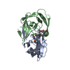
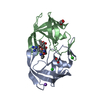
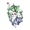
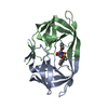
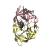
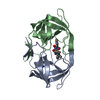
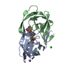
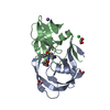
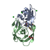
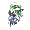
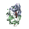
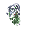
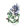
| Title | Cystal structure of HIV-1 Active Site Mutant D25N and p2-NC analog inhibitor | ||||||
 Components Components | Protease (Retropepsin) | ||||||
 Keywords Keywords | HYDROLASE/HYDROLASE INHIBITOR / DRUG RESISTANCE / HIV-1 / HYDROLASE-HYDROLASE INHIBITOR complex | ||||||
| Function / homology |  Function and homology information Function and homology informationHIV-1 retropepsin / symbiont-mediated activation of host apoptosis / retroviral ribonuclease H / exoribonuclease H / exoribonuclease H activity / DNA integration / viral genome integration into host DNA / establishment of integrated proviral latency / RNA-directed DNA polymerase / RNA stem-loop binding ...HIV-1 retropepsin / symbiont-mediated activation of host apoptosis / retroviral ribonuclease H / exoribonuclease H / exoribonuclease H activity / DNA integration / viral genome integration into host DNA / establishment of integrated proviral latency / RNA-directed DNA polymerase / RNA stem-loop binding / viral penetration into host nucleus / host multivesicular body / RNA-directed DNA polymerase activity / RNA-DNA hybrid ribonuclease activity / Transferases; Transferring phosphorus-containing groups; Nucleotidyltransferases / host cell / viral nucleocapsid / DNA recombination / DNA-directed DNA polymerase / aspartic-type endopeptidase activity / Hydrolases; Acting on ester bonds / DNA-directed DNA polymerase activity / symbiont-mediated suppression of host gene expression / viral translational frameshifting / symbiont entry into host cell / lipid binding / host cell nucleus / host cell plasma membrane / virion membrane / structural molecule activity / proteolysis / DNA binding / zinc ion binding Similarity search - Function | ||||||
| Biological species |   Human immunodeficiency virus 1 Human immunodeficiency virus 1 | ||||||
| Method |  X-RAY DIFFRACTION / SYNCHROTRON / AB INITIO / Resolution: 1.05 Å X-RAY DIFFRACTION / SYNCHROTRON / AB INITIO / Resolution: 1.05 Å | ||||||
 Authors Authors | Liu, F. / Weber, I.T. | ||||||
 Citation Citation | Journal: J.Biol.Chem. / Year: 2008 Title: Effect of the Active Site D25N Mutation on the Structure, Stability, and Ligand Binding of the Mature HIV-1 Protease. Authors: Sayer, J.M. / Liu, F. / Ishima, R. / Weber, I.T. / Louis, J.M. #1: Journal: J.Biol.Chem. / Year: 2003Title: Solution structure of the mature HIV-1 protease monomer: insight into the tertiary fold and stability of a precursor. Authors: Ishima, R. / Torchia, D.A. / Lynch, S.M. / Gronenborn, A.M. / Louis, J.M. #2: Journal: J.Mol.Biol. / Year: 2005Title: Kinetic, stability, and structural changes in high-resolution crystal structures of HIV-1 protease with drug-resistant mutations L24I, I50V, and G73S. Authors: Liu, F. / Boross, P.I. / Wang, Y.F. / Tozser, J. / Louis, J.M. / Harrison, R.W. / Weber, I.T. | ||||||
| History |
|
- Structure visualization
| Structure viewer | Molecule: MolmilJmol/JSmol |
|---|
- Downloads & links
-Download
| PDBx/mmCIF format | 3bva.cif.gz | 111.1 KB | Display | PDBx/mmCIF format |
|---|---|---|---|---|
| PDB format | pdb3bva.ent.gz | 84.9 KB | Display | PDB format |
| PDBx/mmJSON format | 3bva.json.gz | Tree view | PDBx/mmJSON format | |
| Others |  Other downloads Other downloads |
-Validation report
| Arichive directory | https://data.pdbj.org/pub/pdb/validation_reports/bv/3bvaftp://data.pdbj.org/pub/pdb/validation_reports/bv/3bva | HTTPS FTP |
|---|
-Related structure data
| Related structure data |  3bvbC  1dazS C: citing same article ( S: Starting model for refinement |
|---|---|
| Similar structure data |

-Links
 PDBj
PDBj


- Assembly
| Deposited unit | 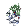
| |||||||||
|---|---|---|---|---|---|---|---|---|---|---|
| 1 |
| |||||||||
| Unit cell |
| |||||||||
| Components on special symmetry positions |
|
-Components
| #1: Protein | Mass: 10739.691 Da / Num. of mol.: 2 / Mutation: Q7K, D25N, L33I, L63I, C67A, C97A Source method: isolated from a genetically manipulated source Source: (gene. exp.) Human immunodeficiency virus 1 / Genus: Lentivirus / Gene: GAG-POL / Production host:  #2: Chemical |   Mass: 92.094 Da / Num. of mol.: 3 / Source method: obtained synthetically / Formula: C3H8O3 Mass: 92.094 Da / Num. of mol.: 3 / Source method: obtained synthetically / Formula: C3H8O3#3: Chemical | ChemComp-2NC / |   Type: peptide-like, Peptide-like / Class: Inhibitor / Mass: 770.983 Da / Num. of mol.: 1 / Source method: obtained synthetically / Formula: C35H68N11O8 Type: peptide-like, Peptide-like / Class: Inhibitor / Mass: 770.983 Da / Num. of mol.: 1 / Source method: obtained synthetically / Formula: C35H68N11O8References: N-{(2S)-2-[(N-acetyl-L-threonyl-L-isoleucyl)amino]hexyl}-L-norleucyl-L-glutaminyl-N~5~-[amino(iminio)methyl]-L-ornithinamide #4: Water | ChemComp-HOH / |  Mass: 18.015 Da / Num. of mol.: 234 / Source method: isolated from a natural source / Formula: H2O Mass: 18.015 Da / Num. of mol.: 234 / Source method: isolated from a natural source / Formula: H2ONonpolymer details | THE INHIBITOR 2NC IS A P2/NC ANALOG. IT HAS A REDUCED PEPTIDE BOND ISOSTERE [CH2-NH] IN PLACE OF ...THE INHIBITOR 2NC IS A P2/NC ANALOG. IT HAS A REDUCED PEPTIDE BOND ISOSTERE [CH2-NH] IN PLACE OF THE SCISSILE AMIDE | |
|---|
-Experimental details
-Experiment
| Experiment | Method: X-RAY DIFFRACTION / Number of used crystals: 1 |
|---|
- Sample preparation
| Crystal | Density Matthews: 2.63 Å3/Da / Density % sol: 54.58 % |
|---|---|
| Crystal grow | Temperature: 295 K / Method: vapor diffusion, hanging drop / pH: 5.4 Details: 10% SODIUM CHLORIDE, CITRATE-PHOSPHATE BUFFER, 7-8% DMSO, pH 5.4, VAPOR DIFFUSION, HANGING DROP, temperature 295K |
-Data collection
| Diffraction | Mean temperature: 95 K |
|---|---|
| Diffraction source | Source: SYNCHROTRON / Site: APS  / Beamline: 22-ID / Wavelength: 1 Å / Beamline: 22-ID / Wavelength: 1 Å |
| Detector | Type: MARRESEARCH / Detector: CCD / Date: Nov 8, 2000 |
| Radiation | Monochromator: SI / Protocol: SINGLE WAVELENGTH / Monochromatic (M) / Laue (L): M / Scattering type: x-ray |
| Radiation wavelength | Wavelength: 1 Å / Relative weight: 1 |
| Reflection | Resolution: 1.05→50 Å / Num. all: 102685 / Num. obs: 97551 / % possible obs: 94.6 % / Observed criterion σ(F): 4 / Observed criterion σ(I): 2 / Redundancy: 7 % / Rmerge(I) obs: 0.136 / Net I/σ(I): 22.3 |
| Reflection shell | Resolution: 1.05→1.09 Å / Redundancy: 2.4 % / Rmerge(I) obs: 0.35 / Mean I/σ(I) obs: 2.7 / % possible all: 82.1 |
- Processing
| Software |
| |||||||||||||||||||||||||||||||||
|---|---|---|---|---|---|---|---|---|---|---|---|---|---|---|---|---|---|---|---|---|---|---|---|---|---|---|---|---|---|---|---|---|---|---|
| Refinement | Method to determine structure: AB INITIO Starting model: PDB entry 1DAZ Resolution: 1.05→10 Å / Num. parameters: 17901 / Num. restraintsaints: 23143 / Cross valid method: FREE R / σ(F): 0 / σ(I): 2 / Stereochemistry target values: ENGH AND HUBER
| |||||||||||||||||||||||||||||||||
| Refine analyze | Num. disordered residues: 37 / Occupancy sum hydrogen: 1663.41 / Occupancy sum non hydrogen: 1775.83 | |||||||||||||||||||||||||||||||||
| Refinement step | Cycle: LAST / Resolution: 1.05→10 Å
| |||||||||||||||||||||||||||||||||
| Refine LS restraints |
|
