 Movie
Movie Controller

[English] 日本語
 Yorodumi
- PDB-3fx5: Structure of HIV-1 Protease in Complex with Potent Inhibitor KNI-... -
+ Open data

- Basic information
| Entry | Database: PDB / ID: 3fx5 | ||||||
|---|---|---|---|---|---|---|---|
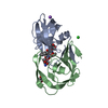
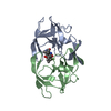
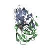
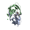
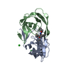
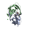
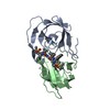
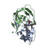
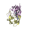
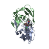
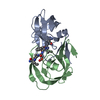
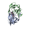
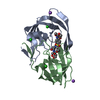
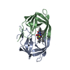
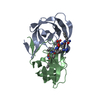
| Title | Structure of HIV-1 Protease in Complex with Potent Inhibitor KNI-272 Determined by High Resolution X-ray Crystallography | ||||||
 Components Components | protease | ||||||
 Keywords Keywords | HYDROLASE/HYDROLASE INHIBITOR / acid protease / homodimer / protease / Hydrolase / Nucleotidyltransferase / Transferase / HYDROLASE-HYDROLASE INHIBITOR complex | ||||||
| Function / homology |  Function and homology information Function and homology informationHIV-1 retropepsin / symbiont-mediated activation of host apoptosis / retroviral ribonuclease H / exoribonuclease H / exoribonuclease H activity / DNA integration / viral genome integration into host DNA / establishment of integrated proviral latency / RNA-directed DNA polymerase / RNA stem-loop binding ...HIV-1 retropepsin / symbiont-mediated activation of host apoptosis / retroviral ribonuclease H / exoribonuclease H / exoribonuclease H activity / DNA integration / viral genome integration into host DNA / establishment of integrated proviral latency / RNA-directed DNA polymerase / RNA stem-loop binding / viral penetration into host nucleus / host multivesicular body / RNA-directed DNA polymerase activity / RNA-DNA hybrid ribonuclease activity / Transferases; Transferring phosphorus-containing groups; Nucleotidyltransferases / host cell / viral nucleocapsid / endonuclease activity / DNA recombination / DNA-directed DNA polymerase / aspartic-type endopeptidase activity / Hydrolases; Acting on ester bonds / DNA-directed DNA polymerase activity / symbiont-mediated suppression of host gene expression / viral translational frameshifting / symbiont entry into host cell / lipid binding / host cell nucleus / host cell plasma membrane / virion membrane / structural molecule activity / proteolysis / DNA binding / zinc ion binding Similarity search - Function | ||||||
| Biological species |   Human immunodeficiency virus 1 Human immunodeficiency virus 1 | ||||||
| Method |  X-RAY DIFFRACTION / SYNCHROTRON / FOURIER SYNTHESIS / Resolution: 0.93 Å X-RAY DIFFRACTION / SYNCHROTRON / FOURIER SYNTHESIS / Resolution: 0.93 Å | ||||||
 Authors Authors | Adachi, M. / Ohhara, T. / Tamada, T. / Okazaki, N. / Kuroki, R. | ||||||
 Citation Citation | Journal: Proc.Natl.Acad.Sci.USA / Year: 2009 Title: Structure of HIV-1 protease in complex with potent inhibitor KNI-272 determined by high-resolution X-ray and neutron crystallography. Authors: Adachi, M. / Ohhara, T. / Kurihara, K. / Tamada, T. / Honjo, E. / Okazaki, N. / Arai, S. / Shoyama, Y. / Kimura, K. / Matsumura, H. / Sugiyama, S. / Adachi, H. / Takano, K. / Mori, Y. / ...Authors: Adachi, M. / Ohhara, T. / Kurihara, K. / Tamada, T. / Honjo, E. / Okazaki, N. / Arai, S. / Shoyama, Y. / Kimura, K. / Matsumura, H. / Sugiyama, S. / Adachi, H. / Takano, K. / Mori, Y. / Hidaka, K. / Kimura, T. / Hayashi, Y. / Kiso, Y. / Kuroki, R. | ||||||
| History |
|
- Structure visualization
| Structure viewer | Molecule: MolmilJmol/JSmol |
|---|
- Downloads & links
-Download
| PDBx/mmCIF format | 3fx5.cif.gz | 163 KB | Display | PDBx/mmCIF format |
|---|---|---|---|---|
| PDB format | pdb3fx5.ent.gz | 132.1 KB | Display | PDB format |
| PDBx/mmJSON format | 3fx5.json.gz | Tree view | PDBx/mmJSON format | |
| Others |  Other downloads Other downloads |
-Validation report
| Arichive directory | https://data.pdbj.org/pub/pdb/validation_reports/fx/3fx5ftp://data.pdbj.org/pub/pdb/validation_reports/fx/3fx5 | HTTPS FTP |
|---|
-Related structure data
| Related structure data |  2zyeC 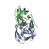 1hpxS S: Starting model for refinement C: citing same article ( |
|---|---|
| Similar structure data |


-Links
 PDBj
PDBj


- Assembly
| Deposited unit | 
| ||||||||||||
|---|---|---|---|---|---|---|---|---|---|---|---|---|---|
| 1 |
| ||||||||||||
| Unit cell |
| ||||||||||||
| Components on special symmetry positions |
|
-Components
| #1: Protein | Mass: 10740.677 Da / Num. of mol.: 2 / Mutation: Q7K, L33I, L63I C67A, C95A Source method: isolated from a genetically manipulated source Source: (gene. exp.) Human immunodeficiency virus 1 / Gene: pol / Plasmid: pET43a / Production host:  References: UniProt: Q9EKL4, UniProt: P03367*PLUS, HIV-1 retropepsin #2: Chemical |   Mass: 92.094 Da / Num. of mol.: 3 / Source method: obtained synthetically / Formula: C3H8O3 Mass: 92.094 Da / Num. of mol.: 3 / Source method: obtained synthetically / Formula: C3H8O3#3: Chemical | ChemComp-KNI / ( |   Type: peptide-like, Peptide-like / Class: Enzyme inhibitor / Mass: 667.839 Da / Num. of mol.: 1 / Source method: obtained synthetically / Formula: C33H41N5O6S2 / References: kynostatin 272 Type: peptide-like, Peptide-like / Class: Enzyme inhibitor / Mass: 667.839 Da / Num. of mol.: 1 / Source method: obtained synthetically / Formula: C33H41N5O6S2 / References: kynostatin 272#4: Water | ChemComp-HOH / |  Mass: 18.015 Da / Num. of mol.: 470 / Source method: isolated from a natural source / Formula: H2O Mass: 18.015 Da / Num. of mol.: 470 / Source method: isolated from a natural source / Formula: H2O |
|---|
-Experimental details
-Experiment
| Experiment | Method: X-RAY DIFFRACTION / Number of used crystals: 1 |
|---|
- Sample preparation
| Crystal | Density Matthews: 2.67 Å3/Da / Density % sol: 53.93 % |
|---|---|
| Crystal grow | Temperature: 293 K / pH: 5 Details: 50mM citrate, 50mM Na-phosphate, 0.061M ammonium sulfate, pH 5.0, temperature 293K |
-Data collection
| Diffraction | Mean temperature: 100 K |
|---|---|
| Diffraction source | Source: SYNCHROTRON / Site: SPring-8  / Beamline: BL41XU / Wavelength: 0.7085 Å / Beamline: BL41XU / Wavelength: 0.7085 Å |
| Detector | Type: ADSC QUANTUM 315 / Detector: CCD / Date: Apr 15, 2007 |
| Radiation | Protocol: SINGLE WAVELENGTH / Monochromatic (M) / Laue (L): M / Scattering type: x-ray |
| Radiation wavelength | Wavelength: 0.7085 Å / Relative weight: 1 |
| Reflection | Resolution: 0.93→47.2 Å / Num. obs: 150216 / % possible obs: 97.2 % / Redundancy: 7.7 % / Rmerge(I) obs: 0.062 / Net I/σ(I): 43.9 |
| Reflection shell | Resolution: 0.93→0.96 Å / Redundancy: 4.1 % / Rmerge(I) obs: 0.295 / Mean I/σ(I) obs: 3.2 / % possible all: 91.8 |
- Processing
| Software |
| ||||||||||||||||||||
|---|---|---|---|---|---|---|---|---|---|---|---|---|---|---|---|---|---|---|---|---|---|
| Refinement | Method to determine structure: FOURIER SYNTHESIS Starting model: PDB ENTRY 1HPX Resolution: 0.93→47.2 Å / σ(F): 0
| ||||||||||||||||||||
| Displacement parameters | Biso max: 125.81 Å2 / Biso min: 3.55 Å2 | ||||||||||||||||||||
| Refinement step | Cycle: LAST / Resolution: 0.93→47.2 Å
| ||||||||||||||||||||
| Refine LS restraints |
|
