- PDB-3bbr: Crystal structure of the iGluR2 ligand binding core (S1S2J-N775S)... -
+
Open data
ID or keywords:
Loading...
-
Basic information
Entry
Database: PDB / ID: 3bbr
Title
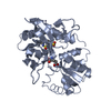
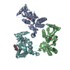
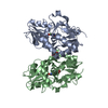
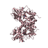
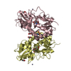
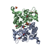
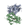
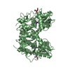
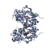
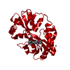
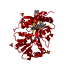
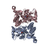
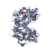
Crystal structure of the iGluR2 ligand binding core (S1S2J-N775S) in complex with a dimeric positive modulator as well as glutamate at 2.25 A resolution
Mass: 18.015 Da / Num. of mol.: 524 / Source method: isolated from a natural source / Formula: H2O
-
Details
Has protein modification
Y
Sequence details
THE NATIVE GLUR2 IS A MEMBRANE PROTEIN. TRANSMEMBRANE REGIONS WERE GENETICALLY REMOVED AND REPLACED ...THE NATIVE GLUR2 IS A MEMBRANE PROTEIN. TRANSMEMBRANE REGIONS WERE GENETICALLY REMOVED AND REPLACED WITH A GLY-THR LINKER.
-
Experimental details
-
Experiment
Experiment
Method: X-RAY DIFFRACTION / Number of used crystals: 1
-
Sample preparation
Crystal
Density Matthews: 2.45 Å3/Da / Density % sol: 49.8 %
Crystal grow
Temperature: 280 K / Method: vapor diffusion, hanging drop / pH: 5.5 Details: 0.3M ammonium sulfate, 0.1M sodium acetate, 25% PEG 4000, pH 5.5, VAPOR DIFFUSION, HANGING DROP, temperature 280K
-
Data collection
Diffraction
Mean temperature: 110 K
Diffraction source
Source: SYNCHROTRON / Site: MAX II / Beamline: I911-2 / Wavelength: 1.043
Detector
Type: MAR CCD 165 mm / Detector: CCD / Date: Mar 13, 2007
Radiation
Protocol: SINGLE WAVELENGTH / Monochromatic (M) / Laue (L): M / Scattering type: x-ray
Radiation wavelength
Wavelength: 1.043 Å / Relative weight: 1
Reflection
Resolution: 2.25→122.169 Å / Num. obs: 27875 / % possible obs: 99.6 % / Redundancy: 6.3 % / Biso Wilson estimate: 16 Å2 / Rmerge(I) obs: 0.09 / Rsym value: 0.09 / Net I/σ(I): 6.7
In the structure databanks used in Yorodumi, some data are registered as the other names, "COVID-19 virus" and "2019-nCoV". Here are the details of the virus and the list of structure data.
Jan 31, 2019. EMDB accession codes are about to change! (news from PDBe EMDB page)
EMDB accession codes are about to change! (news from PDBe EMDB page)
The allocation of 4 digits for EMDB accession codes will soon come to an end. Whilst these codes will remain in use, new EMDB accession codes will include an additional digit and will expand incrementally as the available range of codes is exhausted. The current 4-digit format prefixed with “EMD-” (i.e. EMD-XXXX) will advance to a 5-digit format (i.e. EMD-XXXXX), and so on. It is currently estimated that the 4-digit codes will be depleted around Spring 2019, at which point the 5-digit format will come into force.
The EM Navigator/Yorodumi systems omit the EMD- prefix.
Related info.:Q: What is EMD? / ID/Accession-code notation in Yorodumi/EM Navigator
Yorodumi is a browser for structure data from EMDB, PDB, SASBDB, etc.
This page is also the successor to EM Navigator detail page, and also detail information page/front-end page for Omokage search.
The word "yorodu" (or yorozu) is an old Japanese word meaning "ten thousand". "mi" (miru) is to see.
Related info.:EMDB / PDB / SASBDB / Comparison of 3 databanks / Yorodumi Search / Aug 31, 2016. New EM Navigator & Yorodumi / Yorodumi Papers / Jmol/JSmol / Function and homology information / Changes in new EM Navigator and Yorodumi
 Movie
Movie Controller

 Yorodumi
 Open data
 Basic information
 Components
 Keywords
 Function and homology information
 X-RAY DIFFRACTION /
  Authors
 Citation
 Structure visualization
 Downloads & links
 Other downloads
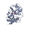


 PDBj
PDBj


 Assembly


 Mass: 96.063 Da / Num. of mol.: 5 / Source method: obtained synthetically / Formula: SO4
 Mass: 35.453 Da / Num. of mol.: 1 / Source method: obtained synthetically / Formula: Cl
 Type: L-peptide linking / Mass: 147.129 Da / Num. of mol.: 2 / Source method: obtained synthetically / Formula: C5H9NO4
 Mass: 424.577 Da / Num. of mol.: 1 / Source method: obtained synthetically / Formula: C20H28N2O4S2
 Mass: 92.094 Da / Num. of mol.: 2 / Source method: obtained synthetically / Formula: C3H8O3
 Sample preparation
 / Beamline: I911-2 / Wavelength: 1.043
  Processing