 Movie
Movie Controller

[English] 日本語
 Yorodumi
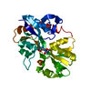
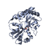
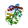
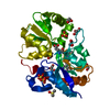
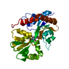
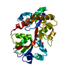
- PDB-1lbc: Crystal structure of GluR2 ligand binding core (S1S2J-N775S) in c... -
+ Open data

- Basic information
| Entry | Database: PDB / ID: 1lbc | ||||||
|---|---|---|---|---|---|---|---|
| Title | Crystal structure of GluR2 ligand binding core (S1S2J-N775S) in complex with cyclothiazide (CTZ) as well as glutamate at 1.8 A resolution | ||||||
 Components Components | Glutamine Receptor 2 | ||||||
 Keywords Keywords | MEMBRANE PROTEIN / AMPA receptor / GluR2 / S1S2 / ligand binding core / point mutation / N775S / cyclothiazide / CTZ / agonist | ||||||
| Function / homology |  Function and homology information Function and homology informationspine synapse / dendritic spine neck / dendritic spine cytoplasm / dendritic spine head / cellular response to amine stimulus / Activation of AMPA receptors / ligand-gated monoatomic cation channel activity / perisynaptic space / Trafficking of GluR2-containing AMPA receptors / response to lithium ion ...spine synapse / dendritic spine neck / dendritic spine cytoplasm / dendritic spine head / cellular response to amine stimulus / Activation of AMPA receptors / ligand-gated monoatomic cation channel activity / perisynaptic space / Trafficking of GluR2-containing AMPA receptors / response to lithium ion / AMPA glutamate receptor activity / AMPA glutamate receptor clustering / kainate selective glutamate receptor activity / immunoglobulin binding / AMPA glutamate receptor complex / regulation of receptor recycling / extracellularly glutamate-gated ion channel activity / cellular response to glycine / ionotropic glutamate receptor complex / asymmetric synapse / Unblocking of NMDA receptors, glutamate binding and activation / glutamate receptor binding / positive regulation of synaptic transmission / conditioned place preference / response to fungicide / regulation of synaptic transmission, glutamatergic / extracellular ligand-gated monoatomic ion channel activity / cytoskeletal protein binding / glutamate-gated receptor activity / cellular response to brain-derived neurotrophic factor stimulus / regulation of long-term synaptic depression / somatodendritic compartment / glutamate-gated calcium ion channel activity / presynaptic active zone membrane / excitatory synapse / ionotropic glutamate receptor signaling pathway / ionotropic glutamate receptor binding / dendrite cytoplasm / dendrite membrane / ligand-gated monoatomic ion channel activity involved in regulation of presynaptic membrane potential / positive regulation of excitatory postsynaptic potential / dendritic shaft / SNARE binding / synaptic membrane / PDZ domain binding / establishment of protein localization / protein tetramerization / synaptic transmission, glutamatergic / transmitter-gated monoatomic ion channel activity involved in regulation of postsynaptic membrane potential / cerebral cortex development / receptor internalization / postsynaptic density membrane / modulation of chemical synaptic transmission / Schaffer collateral - CA1 synapse / terminal bouton / synaptic vesicle / long-term synaptic potentiation / amyloid-beta binding / synaptic vesicle membrane / growth cone / presynapse / signaling receptor activity / presynaptic membrane / scaffold protein binding / dendritic spine / chemical synaptic transmission / perikaryon / postsynaptic membrane / neuron projection / postsynaptic density / external side of plasma membrane / axon / neuronal cell body / synapse / dendrite / protein kinase binding / protein-containing complex binding / glutamatergic synapse / cell surface / endoplasmic reticulum / protein-containing complex / membrane / identical protein binding / plasma membrane Similarity search - Function | ||||||
| Biological species |  | ||||||
| Method |  X-RAY DIFFRACTION / SYNCHROTRON / rigid body refinement / Resolution: 1.8 Å X-RAY DIFFRACTION / SYNCHROTRON / rigid body refinement / Resolution: 1.8 Å | ||||||
 Authors Authors | Sun, Y. / Olson, R. / Horning, M. / Armstrong, N. / Mayer, M. / Gouaux, E. | ||||||
 Citation Citation | Journal: Nature / Year: 2002 Title: Mechanism of glutamate receptor desensitization. Authors: Sun, Y. / Olson, R. / Horning, M. / Armstrong, N. / Mayer, M. / Gouaux, E. | ||||||
| History |
| ||||||
| Remark 999 | Sequence The native GLUR-2 is a membrane protein. The protein crystallized by the author is the ...Sequence The native GLUR-2 is a membrane protein. The protein crystallized by the author is the extracellular ligand binding domain of GLUR-2. Transmembrane regions were genetically removed and replaced with a Gly-Thr linker. The sequence, as a result, matches discontinuously with the reference database. |
- Structure visualization
| Structure viewer | Molecule: MolmilJmol/JSmol |
|---|
- Downloads & links
-Download
| PDBx/mmCIF format | 1lbc.cif.gz | 164.3 KB | Display | PDBx/mmCIF format |
|---|---|---|---|---|
| PDB format | pdb1lbc.ent.gz | 130.7 KB | Display | PDB format |
| PDBx/mmJSON format | 1lbc.json.gz | Tree view | PDBx/mmJSON format | |
| Others |  Other downloads Other downloads |
-Validation report
| Arichive directory | https://data.pdbj.org/pub/pdb/validation_reports/lb/1lbcftp://data.pdbj.org/pub/pdb/validation_reports/lb/1lbc | HTTPS FTP |
|---|
-Related structure data
| Related structure data | 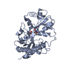 1lb8C 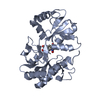 1lb9C  1lbbC  1ftjS S: Starting model for refinement C: citing same article ( |
|---|---|
| Similar structure data |


-Links
 PDBj
PDBj


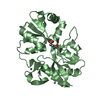
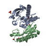
- Assembly
| Deposited unit | 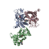
| ||||||||
|---|---|---|---|---|---|---|---|---|---|
| 1 | 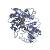
| ||||||||
| 2 | 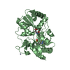
| ||||||||
| 3 | 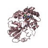
| ||||||||
| 4 | 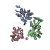
| ||||||||
| 5 | 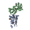
| ||||||||
| Unit cell |
|
-Components
| #1: Protein | Mass: 29194.658 Da / Num. of mol.: 3 / Fragment: ligand binding core / Mutation: N775S Source method: isolated from a genetically manipulated source Source: (gene. exp.)  #2: Chemical | ChemComp-ZN /   Mass: 65.409 Da / Num. of mol.: 5 / Source method: obtained synthetically / Formula: Zn Mass: 65.409 Da / Num. of mol.: 5 / Source method: obtained synthetically / Formula: Zn#3: Chemical |   Type: L-peptide linking / Mass: 147.129 Da / Num. of mol.: 3 / Source method: obtained synthetically / Formula: C5H9NO4 Type: L-peptide linking / Mass: 147.129 Da / Num. of mol.: 3 / Source method: obtained synthetically / Formula: C5H9NO4#4: Chemical | 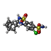  Mass: 389.878 Da / Num. of mol.: 3 / Source method: obtained synthetically / Formula: C14H16ClN3O4S2 Mass: 389.878 Da / Num. of mol.: 3 / Source method: obtained synthetically / Formula: C14H16ClN3O4S2#5: Water | ChemComp-HOH / |  Mass: 18.015 Da / Num. of mol.: 319 / Source method: isolated from a natural source / Formula: H2O Mass: 18.015 Da / Num. of mol.: 319 / Source method: isolated from a natural source / Formula: H2OHas protein modification | Y | |
|---|
-Experimental details
-Experiment
| Experiment | Method: X-RAY DIFFRACTION / Number of used crystals: 1 |
|---|
- Sample preparation
| Crystal | Density Matthews: 2.52 Å3/Da / Density % sol: 51.09 % | |||||||||||||||||||||||||||||||||||
|---|---|---|---|---|---|---|---|---|---|---|---|---|---|---|---|---|---|---|---|---|---|---|---|---|---|---|---|---|---|---|---|---|---|---|---|---|
| Crystal grow | Temperature: 277 K / Method: vapor diffusion, hanging drop / pH: 6.5 Details: pEG 8000 Zn(oAc)2, cacodylate, pH 6.5, VAPOR DIFFUSION, HANGING DROP, temperature 277K | |||||||||||||||||||||||||||||||||||
| Crystal grow | *PLUS Temperature: 4 ℃ | |||||||||||||||||||||||||||||||||||
| Components of the solutions | *PLUS
|
-Data collection
| Diffraction | Mean temperature: 110 K |
|---|---|
| Diffraction source | Source: SYNCHROTRON / Site: NSLS  / Beamline: X4A / Wavelength: 0.9196 Å / Beamline: X4A / Wavelength: 0.9196 Å |
| Detector | Type: ADSC QUANTUM 4 / Detector: CCD / Date: May 20, 2001 |
| Radiation | Protocol: SINGLE WAVELENGTH / Monochromatic (M) / Laue (L): M / Scattering type: x-ray |
| Radiation wavelength | Wavelength: 0.9196 Å / Relative weight: 1 |
| Reflection | Resolution: 1.8→30 Å / Num. all: 81971 / Num. obs: 81971 / % possible obs: 99.6 % / Observed criterion σ(I): -3 |
| Reflection shell | Resolution: 1.8→1.86 Å / % possible all: 98.8 |
| Reflection | *PLUS Lowest resolution: 30 Å / Redundancy: 4.5 % / Rmerge(I) obs: 0.05 |
| Reflection shell | *PLUS % possible obs: 98.8 % / Rmerge(I) obs: 0.346 / Mean I/σ(I) obs: 4 |
- Processing
| Software |
| ||||||||||||||||||||
|---|---|---|---|---|---|---|---|---|---|---|---|---|---|---|---|---|---|---|---|---|---|
| Refinement | Method to determine structure: rigid body refinement Starting model: PDB entry 1FTJ Resolution: 1.8→20 Å / σ(F): 0 / Stereochemistry target values: Engh & Huber
| ||||||||||||||||||||
| Refinement step | Cycle: LAST / Resolution: 1.8→20 Å
| ||||||||||||||||||||
| Refine LS restraints |
| ||||||||||||||||||||
| Refinement | *PLUS Lowest resolution: 30 Å / % reflection Rfree: 10 % / Rfactor obs: 0.222 / Rfactor Rfree: 0.243 / Rfactor Rwork: 0.222 | ||||||||||||||||||||
| Solvent computation | *PLUS | ||||||||||||||||||||
| Displacement parameters | *PLUS |
