 Movie
Movie Controller

[English] 日本語
 Yorodumi
- PDB-1nnp: X-ray structure of the GluR2 ligand-binding core (S1S2J) in compl... -
+ Open data

- Basic information
| Entry | Database: PDB / ID: 1nnp | ||||||
|---|---|---|---|---|---|---|---|
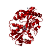
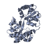
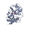
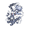
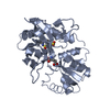
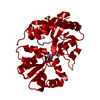
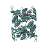
| Title | X-ray structure of the GluR2 ligand-binding core (S1S2J) in complex with (S)-ATPA at 1.9 A resolution. Crystallization without zinc ions. | ||||||
 Components Components | Glutamate receptor 2 | ||||||
 Keywords Keywords | MEMBRANE PROTEIN / Ionotropic glutamate receptor GluR2 / ligand-binding core / agonist complex. | ||||||
| Function / homology |  Function and homology information Function and homology informationspine synapse / dendritic spine neck / dendritic spine cytoplasm / dendritic spine head / cellular response to amine stimulus / Activation of AMPA receptors / ligand-gated monoatomic cation channel activity / perisynaptic space / Trafficking of GluR2-containing AMPA receptors / response to lithium ion ...spine synapse / dendritic spine neck / dendritic spine cytoplasm / dendritic spine head / cellular response to amine stimulus / Activation of AMPA receptors / ligand-gated monoatomic cation channel activity / perisynaptic space / Trafficking of GluR2-containing AMPA receptors / response to lithium ion / AMPA glutamate receptor activity / AMPA glutamate receptor clustering / regulation of receptor recycling / kainate selective glutamate receptor activity / immunoglobulin binding / AMPA glutamate receptor complex / extracellularly glutamate-gated ion channel activity / cellular response to glycine / ionotropic glutamate receptor complex / asymmetric synapse / Unblocking of NMDA receptors, glutamate binding and activation / glutamate receptor binding / positive regulation of synaptic transmission / conditioned place preference / regulation of synaptic transmission, glutamatergic / response to fungicide / extracellular ligand-gated monoatomic ion channel activity / cytoskeletal protein binding / glutamate-gated receptor activity / cellular response to brain-derived neurotrophic factor stimulus / regulation of long-term synaptic depression / somatodendritic compartment / glutamate-gated calcium ion channel activity / presynaptic active zone membrane / ionotropic glutamate receptor signaling pathway / excitatory synapse / ionotropic glutamate receptor binding / dendrite cytoplasm / dendrite membrane / ligand-gated monoatomic ion channel activity involved in regulation of presynaptic membrane potential / positive regulation of excitatory postsynaptic potential / dendritic shaft / SNARE binding / synaptic membrane / PDZ domain binding / protein tetramerization / establishment of protein localization / synaptic transmission, glutamatergic / transmitter-gated monoatomic ion channel activity involved in regulation of postsynaptic membrane potential / receptor internalization / cerebral cortex development / postsynaptic density membrane / modulation of chemical synaptic transmission / Schaffer collateral - CA1 synapse / long-term synaptic potentiation / terminal bouton / synaptic vesicle / amyloid-beta binding / synaptic vesicle membrane / presynapse / growth cone / signaling receptor activity / presynaptic membrane / scaffold protein binding / chemical synaptic transmission / dendritic spine / perikaryon / postsynaptic membrane / neuron projection / postsynaptic density / external side of plasma membrane / axon / neuronal cell body / dendrite / synapse / protein kinase binding / protein-containing complex binding / glutamatergic synapse / cell surface / endoplasmic reticulum / protein-containing complex / membrane / identical protein binding / plasma membrane Similarity search - Function | ||||||
| Biological species |  | ||||||
| Method |  X-RAY DIFFRACTION / SYNCHROTRON / MOLECULAR REPLACEMENT / Resolution: 1.9 Å X-RAY DIFFRACTION / SYNCHROTRON / MOLECULAR REPLACEMENT / Resolution: 1.9 Å | ||||||
 Authors Authors | Lunn, M.L. / Hogner, A. / Stensbol, T.B. / Gouaux, E. / Egebjerg, J. / Kastrup, J.S. | ||||||
 Citation Citation | Journal: J.Med.Chem. / Year: 2003 Title: Three-Dimensional Structure of the Ligand-Binding Core of GluR2 in Complex with the Agonist (S)-ATPA: Implications for Receptor Subunit Selectivity. Authors: Lunn, M.L. / Hogner, A. / Stensbol, T.B. / Gouaux, E. / Egebjerg, J. / Kastrup, J.S. #1: Journal: J.Mol.Biol. / Year: 2002Title: Structural basis for AMPA receptor activation and ligand selectivity: Crystal structures of five agonist complexes with the GluR2 ligand binding core. Authors: Hogner, A. / Kastrup, J.S. / Jin, R. / Liljefors, T. / Mayer, M.L. / Egebjerg, J. / Larsen, I. / Gouaux, E. #2: Journal: Neuron / Year: 2000Title: Mechanisms for activation and antagonism of an AMPA-sensitive glutamate receptor: Crystal structures of the GluR2 ligand binding core. Authors: Armstrong, N. / Gouaux, E. #3: Journal: Nature / Year: 2002Title: Mechanism of glutamate receptor desensitization. Authors: Sun, Y. / Olson, R. / Horning, M. / Armstrong, N. / Mayer, M. / Gouaux, E. #4: Journal: Protein Sci. / Year: 1998Title: Probing the ligand binding domain of the GluR2 receptor by proteolysis and deletion mutagenesis defines domain boundaries and yields a crystallizable construct. Authors: Chen, G.Q. / Sun, R. / Jin, R. / Gouaux, E. | ||||||
| History |
| ||||||
| Remark 300 | BIOMOLECULE: 1 THIS ENTRY CONTAINS THE CRYSTALLOGRAPHIC ASYMMETRIC UNIT WHICH CONSISTS OF 2 ... BIOMOLECULE: 1 THIS ENTRY CONTAINS THE CRYSTALLOGRAPHIC ASYMMETRIC UNIT WHICH CONSISTS OF 2 CHAIN(S). SEE REMARK 350 FOR INFORMATION ON GENERATING THE BIOLOGICAL MOLECULE(S). NOTE THAT COORDINATES FOR ONE DIMER OF A COMPLETE TETRAMERIC MULTIMER REPRESENTING THE KNOWN BIOLOGICALLY SIGNIFICANT OLIGOMERIZATION STATE OF THE MOLECULE CAN BE GENERATED BY APPLYING BIOMT TRANSFORMATIONS GIVEN IN REMARK 350. BOTH NON-CRYSTALLOGRAPHIC AND CRYSTALLOGRAPHIC OPERATIONS ARE GIVEN. | ||||||
| Remark 999 | SEQUENCE Native GluR2 is a membrane protein. The protein crystallized is the extracellular ligand- ...SEQUENCE Native GluR2 is a membrane protein. The protein crystallized is the extracellular ligand-binding core of GluR2. Transmembrane regions were genetically removed and replaced with a Gly-Thr linker (residues 115-116). Therefore, the sequence matches discontinuously with the reference database (413-527, 653-796). The two first residues of the sequence (Gly-2, Ala-1) are cloning artifacts and were not located in the electron density map. |
- Structure visualization
| Structure viewer | Molecule: MolmilJmol/JSmol |
|---|
- Downloads & links
-Download
| PDBx/mmCIF format | 1nnp.cif.gz | 133.4 KB | Display | PDBx/mmCIF format |
|---|---|---|---|---|
| PDB format | pdb1nnp.ent.gz | 102.1 KB | Display | PDB format |
| PDBx/mmJSON format | 1nnp.json.gz | Tree view | PDBx/mmJSON format | |
| Others |  Other downloads Other downloads |
-Validation report
| Arichive directory | https://data.pdbj.org/pub/pdb/validation_reports/nn/1nnpftp://data.pdbj.org/pub/pdb/validation_reports/nn/1nnp | HTTPS FTP |
|---|
-Related structure data


-Links
 PDBj
PDBj


- Assembly
| Deposited unit | 
| ||||||||
|---|---|---|---|---|---|---|---|---|---|
| 1 |
| ||||||||
| Unit cell |
| ||||||||
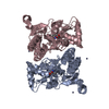
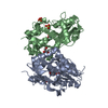
| Details | The biological assembly is a tetramer composed of dimers-of-dimers. Only the dimer is observed in the crystal. The dimer may be generated by applying the following to chain B: TRANSFORM FRACTIONAL - 1.00000 0.00000 0.00000 - 0.00000 -1.00000 0.00000 - 0.00000 0.00000 -1.00000 - -0.50000 0.50000 1.00000 |
-Components
| #1: Protein | Mass: 29221.682 Da / Num. of mol.: 2 / Fragment: GluR2-flop ligand-binding core (S1S2J) Source method: isolated from a genetically manipulated source Source: (gene. exp.)  #2: Chemical |   Mass: 96.063 Da / Num. of mol.: 2 / Source method: obtained synthetically / Formula: SO4 Mass: 96.063 Da / Num. of mol.: 2 / Source method: obtained synthetically / Formula: SO4#3: Chemical |   Mass: 227.237 Da / Num. of mol.: 2 / Source method: obtained synthetically / Formula: C10H15N2O4 Mass: 227.237 Da / Num. of mol.: 2 / Source method: obtained synthetically / Formula: C10H15N2O4#4: Water | ChemComp-HOH / |  Mass: 18.015 Da / Num. of mol.: 725 / Source method: isolated from a natural source / Formula: H2O Mass: 18.015 Da / Num. of mol.: 725 / Source method: isolated from a natural source / Formula: H2OHas protein modification | Y | |
|---|
-Experimental details
-Experiment
| Experiment | Method: X-RAY DIFFRACTION / Number of used crystals: 1 |
|---|
- Sample preparation
| Crystal | Density Matthews: 2.4 Å3/Da / Density % sol: 48 % | ||||||||||||||||||||||||||||||||||||
|---|---|---|---|---|---|---|---|---|---|---|---|---|---|---|---|---|---|---|---|---|---|---|---|---|---|---|---|---|---|---|---|---|---|---|---|---|---|
| Crystal grow | Temperature: 279 K / Method: vapor diffusion, hanging drop / pH: 5.6 Details: ammonium sulfate, PEG8000, sodium acetate, pH 5.6, VAPOR DIFFUSION, HANGING DROP, temperature 279K | ||||||||||||||||||||||||||||||||||||
| Crystal grow | *PLUS Temperature: 6 ℃ | ||||||||||||||||||||||||||||||||||||
| Components of the solutions | *PLUS
|
-Data collection
| Diffraction | Mean temperature: 100 K |
|---|---|
| Diffraction source | Source: SYNCHROTRON / Site: MAX II  / Beamline: I711 / Wavelength: 1.076 Å / Beamline: I711 / Wavelength: 1.076 Å |
| Detector | Type: MARRESEARCH / Detector: CCD / Date: Apr 25, 2002 |
| Radiation | Protocol: SINGLE WAVELENGTH / Monochromatic (M) / Laue (L): M / Scattering type: x-ray |
| Radiation wavelength | Wavelength: 1.076 Å / Relative weight: 1 |
| Reflection | Resolution: 1.9→20 Å / Num. all: 40611 / Num. obs: 40611 / % possible obs: 92.2 % / Observed criterion σ(F): 0 / Observed criterion σ(I): 0 / Redundancy: 4.1 % / Biso Wilson estimate: 13.5 Å2 / Rmerge(I) obs: 0.092 / Net I/σ(I): 13.1 |
| Reflection shell | Resolution: 1.9→1.97 Å / Rmerge(I) obs: 0.348 / Mean I/σ(I) obs: 2.4 / Num. unique all: 3546 / % possible all: 81.6 |
| Reflection | *PLUS Lowest resolution: 20 Å |
| Reflection shell | *PLUS % possible obs: 81.6 % |
- Processing
| Software |
| ||||||||||||||||||||||||||||||||||||
|---|---|---|---|---|---|---|---|---|---|---|---|---|---|---|---|---|---|---|---|---|---|---|---|---|---|---|---|---|---|---|---|---|---|---|---|---|---|
| Refinement | Method to determine structure: MOLECULAR REPLACEMENT Starting model: GluR2:(S)-ATPA complex (zinc form). Resolution: 1.9→20 Å / Rfactor Rfree error: 0.005 / Data cutoff high rms absF: 2164674.72 / Isotropic thermal model: RESTRAINED / Cross valid method: THROUGHOUT / σ(F): 0 / Stereochemistry target values: Engh & Huber Details: The first three N-terminal residues and the last two C-terminal residues were not located in the electron density map.
| ||||||||||||||||||||||||||||||||||||
| Solvent computation | Bsol: 48.6949 Å2 / ksol: 0.360816 e/Å3 | ||||||||||||||||||||||||||||||||||||
| Displacement parameters | Biso mean: 22 Å2
| ||||||||||||||||||||||||||||||||||||
| Refine analyze |
| ||||||||||||||||||||||||||||||||||||
| Refinement step | Cycle: LAST / Resolution: 1.9→20 Å
| ||||||||||||||||||||||||||||||||||||
| Refine LS restraints |
| ||||||||||||||||||||||||||||||||||||
| LS refinement shell | Resolution: 1.9→2.02 Å / Rfactor Rfree error: 0.016 / Total num. of bins used: 6
| ||||||||||||||||||||||||||||||||||||
| Xplor file | Serial no: 1 / Param file: protein_rep.param / Topol file: protein.top | ||||||||||||||||||||||||||||||||||||
| Refinement | *PLUS Lowest resolution: 20 Å / % reflection Rfree: 4.6 % / Rfactor Rfree: 0.217 / Rfactor Rwork: 0.19 | ||||||||||||||||||||||||||||||||||||
| Solvent computation | *PLUS | ||||||||||||||||||||||||||||||||||||
| Displacement parameters | *PLUS | ||||||||||||||||||||||||||||||||||||
| Refine LS restraints | *PLUS
|
