 Movie
Movie Controller

[English] 日本語
 Yorodumi
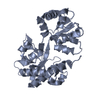
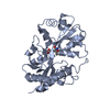
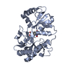
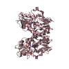
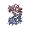
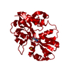
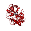
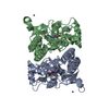
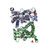
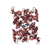
- PDB-1m5e: X-RAY STRUCTURE OF THE GLUR2 LIGAND BINDING CORE (S1S2J) IN COMPL... -
+ Open data

- Basic information
| Entry | Database: PDB / ID: 1m5e | ||||||
|---|---|---|---|---|---|---|---|
| Title | X-RAY STRUCTURE OF THE GLUR2 LIGAND BINDING CORE (S1S2J) IN COMPLEX WITH ACPA AT 1.46 A RESOLUTION | ||||||
 Components Components | Glutamate receptor 2 | ||||||
 Keywords Keywords | MEMBRANE PROTEIN / Ionotropic glutamate receptor / GluR2 / ligand binding core / agonist complex. | ||||||
| Function / homology |  Function and homology information Function and homology informationspine synapse / dendritic spine neck / dendritic spine cytoplasm / dendritic spine head / cellular response to amine stimulus / Activation of AMPA receptors / ligand-gated monoatomic cation channel activity / perisynaptic space / Trafficking of GluR2-containing AMPA receptors / response to lithium ion ...spine synapse / dendritic spine neck / dendritic spine cytoplasm / dendritic spine head / cellular response to amine stimulus / Activation of AMPA receptors / ligand-gated monoatomic cation channel activity / perisynaptic space / Trafficking of GluR2-containing AMPA receptors / response to lithium ion / AMPA glutamate receptor activity / AMPA glutamate receptor clustering / kainate selective glutamate receptor activity / immunoglobulin binding / AMPA glutamate receptor complex / regulation of receptor recycling / extracellularly glutamate-gated ion channel activity / cellular response to glycine / ionotropic glutamate receptor complex / asymmetric synapse / Unblocking of NMDA receptors, glutamate binding and activation / glutamate receptor binding / conditioned place preference / positive regulation of synaptic transmission / response to fungicide / regulation of synaptic transmission, glutamatergic / extracellular ligand-gated monoatomic ion channel activity / cytoskeletal protein binding / glutamate-gated receptor activity / cellular response to brain-derived neurotrophic factor stimulus / regulation of long-term synaptic depression / somatodendritic compartment / glutamate-gated calcium ion channel activity / presynaptic active zone membrane / excitatory synapse / ionotropic glutamate receptor signaling pathway / ionotropic glutamate receptor binding / dendrite membrane / dendrite cytoplasm / ligand-gated monoatomic ion channel activity involved in regulation of presynaptic membrane potential / positive regulation of excitatory postsynaptic potential / dendritic shaft / SNARE binding / synaptic membrane / PDZ domain binding / establishment of protein localization / protein tetramerization / transmitter-gated monoatomic ion channel activity involved in regulation of postsynaptic membrane potential / synaptic transmission, glutamatergic / cerebral cortex development / receptor internalization / postsynaptic density membrane / modulation of chemical synaptic transmission / Schaffer collateral - CA1 synapse / terminal bouton / synaptic vesicle / long-term synaptic potentiation / amyloid-beta binding / synaptic vesicle membrane / growth cone / presynapse / signaling receptor activity / scaffold protein binding / presynaptic membrane / dendritic spine / chemical synaptic transmission / perikaryon / postsynaptic membrane / neuron projection / postsynaptic density / external side of plasma membrane / axon / neuronal cell body / synapse / dendrite / protein kinase binding / protein-containing complex binding / glutamatergic synapse / cell surface / endoplasmic reticulum / protein-containing complex / membrane / identical protein binding / plasma membrane Similarity search - Function | ||||||
| Biological species |  | ||||||
| Method |  X-RAY DIFFRACTION / SYNCHROTRON / Difference Fourier / Resolution: 1.46 Å X-RAY DIFFRACTION / SYNCHROTRON / Difference Fourier / Resolution: 1.46 Å | ||||||
 Authors Authors | Hogner, A. / Kastrup, J.S. / Jin, R. / Liljefors, T. / Mayer, M.L. / Egebjerg, J. / Larsen, I.K. / Gouaux, E. | ||||||
 Citation Citation | Journal: J.Mol.Biol. / Year: 2002 Title: Structural Basis for AMPA Receptor Activation and Ligand Selectivity: Crystal Structures of Five Agonist Complexes with the GluR2 Ligand-binding Core Authors: Hogner, A. / Kastrup, J.S. / Jin, R. / Liljefors, T. / Mayer, M.L. / Egebjerg, J. / Larsen, I.K. / Gouaux, E. #1: Journal: Neuron / Year: 2000Title: Mechanisms for activation and antagonism of an AMPA-sensitive glutamate receptor: Crystal structures of the GluR2 ligand binding core. Authors: Armstrong, N. / Gouaux, E. #2: Journal: Nature / Year: 2002Title: Mechanism of glutamate receptor desensitization. Authors: Sun, Y. / Olson, R. / Horning, M. / Armstrong, N. / Mayer, M. / Gouaux, E. #3: Journal: Protein Sci. / Year: 1998Title: Probing the ligand binding domain of the GluR2 receptor by proteolysis and deletion mutagenesis defines domain boundaries and yields a crystallizable construct. Authors: Chen, G.Q. / Sun, Y. / Jin, R. / Gouaux, E. | ||||||
| History |
| ||||||
| Remark 999 | Sequence Native GluR2 is a membrane protein. The protein crystallized is the extracellular ligand ...Sequence Native GluR2 is a membrane protein. The protein crystallized is the extracellular ligand binding domain of GluR2. Transmembrane regions were genetically removed and replaced with a GLY-THR linker (residues 118 and 119). Therefore, the sequence matches discontinuously with the reference database (413-527, 653-796). Residues GLY1 and ALA2 are cloning artifacts. |
- Structure visualization
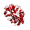
| Structure viewer | Molecule: MolmilJmol/JSmol |
|---|
- Downloads & links
-Download
| PDBx/mmCIF format | 1m5e.cif.gz | 187.3 KB | Display | PDBx/mmCIF format |
|---|---|---|---|---|
| PDB format | pdb1m5e.ent.gz | 148 KB | Display | PDB format |
| PDBx/mmJSON format | 1m5e.json.gz | Tree view | PDBx/mmJSON format | |
| Others |  Other downloads Other downloads |
-Validation report
| Arichive directory | https://data.pdbj.org/pub/pdb/validation_reports/m5/1m5eftp://data.pdbj.org/pub/pdb/validation_reports/m5/1m5e | HTTPS FTP |
|---|
-Related structure data
| Related structure data |  1m5bSC  1m5cC 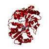 1m5dC  1m5fC S: Starting model for refinement C: citing same article ( |
|---|---|
| Similar structure data |


-Links
 PDBj
PDBj


- Assembly
| Deposited unit | 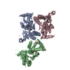
| ||||||||
|---|---|---|---|---|---|---|---|---|---|
| 1 | 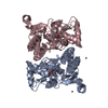
| ||||||||
| 2 | 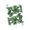
| ||||||||
| 3 | 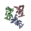
| ||||||||
| Unit cell |
| ||||||||
| Components on special symmetry positions |
| ||||||||
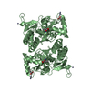
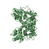
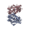
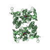
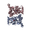
| Details | The biological assembly is a dimer. Chain A and chain C of the asymmetric unit form a non-crystallographic dimer. The dimer of chain B can be generated by the two fold axis: -x, -y, z. |
-Components
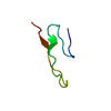
| #1: Protein | Mass: 29221.682 Da / Num. of mol.: 3 / Fragment: GluR2-flop ligand binding core (S1S2J) Source method: isolated from a genetically manipulated source Source: (gene. exp.)  #2: Chemical | ChemComp-ZN /   Mass: 65.409 Da / Num. of mol.: 8 / Source method: obtained synthetically / Formula: Zn Mass: 65.409 Da / Num. of mol.: 8 / Source method: obtained synthetically / Formula: Zn#3: Chemical | 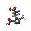  Mass: 214.175 Da / Num. of mol.: 3 / Source method: obtained synthetically / Formula: C8H10N2O5 Mass: 214.175 Da / Num. of mol.: 3 / Source method: obtained synthetically / Formula: C8H10N2O5#4: Chemical | ChemComp-ACT / |   Mass: 59.044 Da / Num. of mol.: 1 / Source method: obtained synthetically / Formula: C2H3O2 Mass: 59.044 Da / Num. of mol.: 1 / Source method: obtained synthetically / Formula: C2H3O2#5: Water | ChemComp-HOH / |  Mass: 18.015 Da / Num. of mol.: 979 / Source method: isolated from a natural source / Formula: H2O Mass: 18.015 Da / Num. of mol.: 979 / Source method: isolated from a natural source / Formula: H2OHas protein modification | Y | |
|---|
-Experimental details
-Experiment
| Experiment | Method: X-RAY DIFFRACTION / Number of used crystals: 1 |
|---|
- Sample preparation
| Crystal | Density Matthews: 2.49 Å3/Da / Density % sol: 0.506 % | |||||||||||||||||||||||||||||||||||||||||||||||||||||||||||||||
|---|---|---|---|---|---|---|---|---|---|---|---|---|---|---|---|---|---|---|---|---|---|---|---|---|---|---|---|---|---|---|---|---|---|---|---|---|---|---|---|---|---|---|---|---|---|---|---|---|---|---|---|---|---|---|---|---|---|---|---|---|---|---|---|---|
| Crystal grow | Temperature: 279 K / Method: vapor diffusion, hanging drop / pH: 6.5 Details: PEG 8000, Zn(OAc)2, cacodylate, pH 6.5, VAPOR DIFFUSION, HANGING DROP, temperature 279K | |||||||||||||||||||||||||||||||||||||||||||||||||||||||||||||||
| Crystal grow | *PLUS Temperature: 4 ℃ / pH: 7 | |||||||||||||||||||||||||||||||||||||||||||||||||||||||||||||||
| Components of the solutions | *PLUS
|
-Data collection
| Diffraction | Mean temperature: 110 K |
|---|---|
| Diffraction source | Source: SYNCHROTRON / Site: EMBL/DESY, HAMBURG  / Beamline: BW7B / Wavelength: 0.842 Å / Beamline: BW7B / Wavelength: 0.842 Å |
| Detector | Type: MARRESEARCH / Detector: IMAGE PLATE / Date: Aug 12, 2000 |
| Radiation | Protocol: SINGLE WAVELENGTH / Monochromatic (M) / Laue (L): M / Scattering type: x-ray |
| Radiation wavelength | Wavelength: 0.842 Å / Relative weight: 1 |
| Reflection | Resolution: 1.46→20 Å / Num. all: 148394 / Num. obs: 148394 / % possible obs: 97.4 % / Observed criterion σ(I): -3 / Redundancy: 4.2 % / Biso Wilson estimate: 16.1 Å2 / Rmerge(I) obs: 0.045 / Net I/σ(I): 20.8 |
| Reflection shell | Resolution: 1.46→1.49 Å / Rmerge(I) obs: 0.416 / Mean I/σ(I) obs: 2.5 / Num. unique all: 7526 / % possible all: 93.9 |
| Reflection | *PLUS Lowest resolution: 20 Å |
| Reflection shell | *PLUS % possible obs: 93.9 % |
- Processing
| Software |
| ||||||||||||||||||||||||||||||||||||
|---|---|---|---|---|---|---|---|---|---|---|---|---|---|---|---|---|---|---|---|---|---|---|---|---|---|---|---|---|---|---|---|---|---|---|---|---|---|
| Refinement | Method to determine structure: Difference Fourier Starting model: PDB entry 1M5B (S1S2J:2-Me-Tet-AMPA, molecule A). Resolution: 1.46→16.75 Å / Rfactor Rfree error: 0.003 / Data cutoff high absF: 2045331 / Data cutoff high rms absF: 2045331 / Isotropic thermal model: RESTRAINED / Cross valid method: THROUGHOUT / σ(F): 0 / Stereochemistry target values: Engh & Huber Details: Residues 1-3 and 262-263 were not located in the electron density map. The side chains of the following residues are not fully defined: LYS A21, LYS A45, LYS A50, ASP A67, THR A131, LYS ...Details: Residues 1-3 and 262-263 were not located in the electron density map. The side chains of the following residues are not fully defined: LYS A21, LYS A45, LYS A50, ASP A67, THR A131, LYS A151, GLU B27, ASP B67, GLU B132, LYS B151, LYS C21, GLU C24, GLU C27, ARG C64, GLU C122, THR C131, GLU C132, ARG C172, LYS C185
| ||||||||||||||||||||||||||||||||||||
| Solvent computation | Solvent model: FLAT MODEL / Bsol: 54.062 Å2 / ksol: 0.437671 e/Å3 | ||||||||||||||||||||||||||||||||||||
| Displacement parameters | Biso mean: 20.3 Å2
| ||||||||||||||||||||||||||||||||||||
| Refine analyze |
| ||||||||||||||||||||||||||||||||||||
| Refinement step | Cycle: LAST / Resolution: 1.46→16.75 Å
| ||||||||||||||||||||||||||||||||||||
| Refine LS restraints |
| ||||||||||||||||||||||||||||||||||||
| LS refinement shell | Resolution: 1.46→1.55 Å / Rfactor Rfree error: 0.008 / Total num. of bins used: 6
| ||||||||||||||||||||||||||||||||||||
| Xplor file | Serial no: 1 / Param file: PROTEIN_REP.PARAM / Topol file: PROTEIN.TOP | ||||||||||||||||||||||||||||||||||||
| Refinement | *PLUS Lowest resolution: 20 Å / % reflection Rfree: 5 % | ||||||||||||||||||||||||||||||||||||
| Solvent computation | *PLUS | ||||||||||||||||||||||||||||||||||||
| Displacement parameters | *PLUS | ||||||||||||||||||||||||||||||||||||
| Refine LS restraints | *PLUS
| ||||||||||||||||||||||||||||||||||||
| LS refinement shell | *PLUS Rfactor Rfree: 0.26 |
