 ムービー
ムービー コントローラー


+ データを開く

- 基本情報
基本情報
| 登録情報 | データベース: PDB / ID: 3bat | ||||||
|---|---|---|---|---|---|---|---|
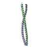
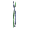
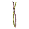
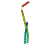
| タイトル | Crystal structure of the N-terminal region of the scallop myosin rod, monoclinic (P21) form | ||||||
 要素 要素 | Myosin heavy chain, striated muscle/General control protein GCN4 | ||||||
 キーワード キーワード | CONTRACTILE PROTEIN / Alpha-helical coiled coil / disorder / salt links / Actin-binding / ATP-binding / Calmodulin-binding / Cytoplasm / Motor protein / Muscle protein / Myosin / Nucleotide-binding / Thick filament | ||||||
| 機能・相同性 |  機能・相同性情報 機能・相同性情報myosin filament organization / myofibril assembly / FCERI mediated MAPK activation / protein localization to nuclear periphery / Activation of the AP-1 family of transcription factors / negative regulation of ribosomal protein gene transcription by RNA polymerase II / response to amino acid starvation / positive regulation of cellular response to amino acid starvation / mediator complex binding / myosin filament ...myosin filament organization / myofibril assembly / FCERI mediated MAPK activation / protein localization to nuclear periphery / Activation of the AP-1 family of transcription factors / negative regulation of ribosomal protein gene transcription by RNA polymerase II / response to amino acid starvation / positive regulation of cellular response to amino acid starvation / mediator complex binding / myosin filament / Oxidative Stress Induced Senescence / locomotion / myosin II complex / structural constituent of muscle / microfilament motor activity / myofibril / amino acid biosynthetic process / TFIID-class transcription factor complex binding / positive regulation of RNA polymerase II transcription preinitiation complex assembly / positive regulation of transcription initiation by RNA polymerase II / cellular response to nutrient levels / muscle contraction / cellular response to amino acid starvation / RNA polymerase II transcription regulator complex / actin filament binding / DNA-binding transcription activator activity, RNA polymerase II-specific / transcription regulator complex / sequence-specific DNA binding / RNA polymerase II-specific DNA-binding transcription factor binding / DNA-binding transcription factor activity, RNA polymerase II-specific / calmodulin binding / intracellular signal transduction / RNA polymerase II cis-regulatory region sequence-specific DNA binding / DNA-binding transcription factor activity / chromatin binding / negative regulation of transcription by RNA polymerase II / positive regulation of transcription by RNA polymerase II / ATP binding / identical protein binding / nucleus 類似検索 - 分子機能 | ||||||
| 生物種 |  Argopecten irradians (無脊椎動物) Argopecten irradians (無脊椎動物) | ||||||
| 手法 |  X線回折 / シンクロトロン / 分子置換 / 解像度: 2.3 Å X線回折 / シンクロトロン / 分子置換 / 解像度: 2.3 Å | ||||||
 データ登録者 データ登録者 | Brown, J.H. / Cohen, C. | ||||||
 引用 引用 | ジャーナル: J.Mol.Biol. / 年: 2008 タイトル: An unstable head-rod junction may promote folding into the compact off-state conformation of regulated myosins. 著者: Brown, J.H. / Yang, Y. / Reshetnikova, L. / Gourinath, S. / Suveges, D. / Kardos, J. / Hobor, F. / Reutzel, R. / Nyitray, L. / Cohen, C. #1: ジャーナル: Nature / 年: 2003タイトル: Visualization of an unstable coiled coil from the scallop myosin rod. 著者: Li, Y. / Brown, J.H. / Reshetnikova, L. / Blazsek, A. / Farkas, L. / Nyitray, L. / Cohen, C. #2: ジャーナル: Proc Natl Acad Sci U S A / 年: 2006タイトル: Crystal structures of human cardiac beta-myosin II S2-Delta provide insight into the functional role of the S2 subfragment. 著者: Wulf Blankenfeldt / Nicolas H Thomä / John S Wray / Mathias Gautel / Ilme Schlichting /  要旨: Myosin II is the major component of the muscle thick filament. It consists of two N-terminal S1 subfragments ("heads") connected to a long dimeric coiled-coil rod. The rod is in itself twofold ...Myosin II is the major component of the muscle thick filament. It consists of two N-terminal S1 subfragments ("heads") connected to a long dimeric coiled-coil rod. The rod is in itself twofold symmetric, but in the filament, the two heads point away from the filament surface and are therefore not equivalent. This breaking of symmetry requires the initial section of the rod, subfragment 2 (S2), to be relatively flexible. S2 is an important functional element, involved in various mechanisms by which the activity of smooth and striated muscle is regulated. We have determined crystal structures of the 126 N-terminal residues of S2 from human cardiac beta-myosin II (S2-Delta), of both WT and the disease-associated E924K mutant. S2-Delta is a straight parallel dimeric coiled coil, but the N terminus of one chain is disordered in WT-S2-Delta due to crystal contacts, indicative of unstable local structure. Bulky noncanonical side chains pack into a/d positions of S2-Delta's N terminus, leading to defined local asymmetry and axial stagger, which could induce nonequivalence of the S1 subfragments. Additionally, S2 possesses a conserved charge distribution with three prominent rings of negative potential within S2-Delta, the first of which may provide a binding interface for the "blocked head" of smooth muscle myosin in the OFF state. The observation that many disease-associated mutations affect the second negatively charged ring further suggests that charge interactions play an important role in regulation of cardiac muscle activity through myosin-binding protein C. | ||||||
| 履歴 |
| ||||||
| Remark 999 | SEQUENCE THE PEPTIDE CONTAINS A GSHM TETRAPEPTIDE, FOLLOWED BY THE N-TERMINAL 51 RESIDUES OF THE ... SEQUENCE THE PEPTIDE CONTAINS A GSHM TETRAPEPTIDE, FOLLOWED BY THE N-TERMINAL 51 RESIDUES OF THE BAY SCALLOP MYOSIN ROD (RESIDUES 835-885 OF THE BAY SCALLOP MYOSIN HEAVY CHAIN, GI:5612), FOLLOWED BY A GS LINKER, FOLLOWED BY THE LEUCINE ZIPPER OF THE YEAST GCN4 TRANSCRIPTION FACTOR (RESIDUES 250-281 OF GI:171584 DENOTED AS RESIDUES 888-919 IN THE SUBMITTED COORDINATES). |
- 構造の表示
構造の表示
| 構造ビューア | 分子: MolmilJmol/JSmol |
|---|
- ダウンロードとリンク
-ダウンロード
| PDBx/mmCIF形式 | 3bat.cif.gz | 80.2 KB | 表示 | PDBx/mmCIF形式 |
|---|---|---|---|---|
| PDB形式 | pdb3bat.ent.gz | 61.9 KB | 表示 | PDB形式 |
| PDBx/mmJSON形式 | 3bat.json.gz | ツリー表示 | PDBx/mmJSON形式 | |
| その他 |  その他のダウンロード その他のダウンロード |
-検証レポート
| アーカイブディレクトリ | https://data.pdbj.org/pub/pdb/validation_reports/ba/3batftp://data.pdbj.org/pub/pdb/validation_reports/ba/3bat | HTTPS FTP |
|---|
-関連構造データ


-リンク
 PDBj
PDBj


- 集合体
集合体
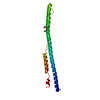
| 登録構造単位 | 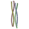
| ||||||||
|---|---|---|---|---|---|---|---|---|---|
| 1 | 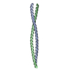
| ||||||||
| 2 | 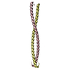
| ||||||||
| 単位格子 |
|
-要素
| #1: タンパク質 | 分子量: 10559.174 Da / 分子数: 4 断片: Bay Scallop Myosin (Residues 835-885)/Yeast GCN4 Transcription Factor (Residues 250-281) 由来タイプ: 組換発現 由来: (組換発現) Argopecten irradians (無脊椎動物), (組換発現) 属: Argopecten, Saccharomyces / 生物種: , / 株: , / 組織: Adductor muscle/- / 遺伝子: -/GCN4, AAS3, ARG9, YEL009C / プラスミド: pET15b / 発現宿主:  #2: 水 | ChemComp-HOH / |  分子量: 18.015 Da / 分子数: 99 / 由来タイプ: 天然 / 式: H2O 分子量: 18.015 Da / 分子数: 99 / 由来タイプ: 天然 / 式: H2O |
|---|
-実験情報
-実験
| 実験 | 手法: X線回折 / 使用した結晶の数: 1 |
|---|
- 試料調製
試料調製
| 結晶 | マシュー密度: 2.25 Å3/Da / 溶媒含有率: 45.45 % |
|---|---|
| 結晶化 | 温度: 277 K / 手法: 蒸気拡散法, ハンギングドロップ法 / pH: 6.2 詳細: 2 microliters of protein solution (4 mg/ml protein in 30 mM MOPS buffer pH 7.2, 40 mM NaCl, 2 mM NaN3) mixed with 2 microliters of (25% PEG 3350, 50 mM NH4I) and equilibrated against 1 ml of ...詳細: 2 microliters of protein solution (4 mg/ml protein in 30 mM MOPS buffer pH 7.2, 40 mM NaCl, 2 mM NaN3) mixed with 2 microliters of (25% PEG 3350, 50 mM NH4I) and equilibrated against 1 ml of (17.5% PEG 3350, 35 mM NH4I, 28 mM NaCl, 2 mM NaN3, 20 mM MOPS pH 6.2). Harvested crystals were cryoprotected in 25.5% PEG 3350 and 15% glycerol, VAPOR DIFFUSION, HANGING DROP, temperature 277K |
-データ収集
| 回折 | 平均測定温度: 100 K | |||||||||||||||||||||||||||||||||||||||||||||||||||||||
|---|---|---|---|---|---|---|---|---|---|---|---|---|---|---|---|---|---|---|---|---|---|---|---|---|---|---|---|---|---|---|---|---|---|---|---|---|---|---|---|---|---|---|---|---|---|---|---|---|---|---|---|---|---|---|---|---|
| 放射光源 | 由来: シンクロトロン / サイト: NSLS  / ビームライン: X26C / 波長: 0.9791 Å / ビームライン: X26C / 波長: 0.9791 Å | |||||||||||||||||||||||||||||||||||||||||||||||||||||||
| 検出器 | タイプ: ADSC QUANTUM 4 / 検出器: CCD 詳細: channel-cut Si(111) crystal monochromator followed by a doubly focusing toroidal mirror | |||||||||||||||||||||||||||||||||||||||||||||||||||||||
| 放射 | モノクロメーター: Si(111) crystal / プロトコル: SINGLE WAVELENGTH / 単色(M)・ラウエ(L): M / 散乱光タイプ: x-ray | |||||||||||||||||||||||||||||||||||||||||||||||||||||||
| 放射波長 | 波長: 0.9791 Å / 相対比: 1 | |||||||||||||||||||||||||||||||||||||||||||||||||||||||
| 反射 | 解像度: 1.95→30 Å / Num. obs: 27340 / % possible obs: 98.5 % / Rmerge(I) obs: 0.052 / Χ2: 1.085 / Net I/σ(I): 13.8 | |||||||||||||||||||||||||||||||||||||||||||||||||||||||
| 反射 シェル |
|
- 解析
解析
| ソフトウェア |
| ||||||||||||||||||||||||
|---|---|---|---|---|---|---|---|---|---|---|---|---|---|---|---|---|---|---|---|---|---|---|---|---|---|
| 精密化 | 構造決定の手法: 分子置換 開始モデル: PDB entry 1NKN 解像度: 2.3→30 Å / 交差検証法: THROUGHOUT / σ(F): 0
| ||||||||||||||||||||||||
| 溶媒の処理 | Bsol: 38.499 Å2 | ||||||||||||||||||||||||
| 原子変位パラメータ | Biso mean: 53.827 Å2
| ||||||||||||||||||||||||
| Refine analyze |
| ||||||||||||||||||||||||
| 精密化ステップ | サイクル: LAST / 解像度: 2.3→30 Å
| ||||||||||||||||||||||||
| 拘束条件 |
| ||||||||||||||||||||||||
| LS精密化 シェル | 解像度: 2.3→2.44 Å
| ||||||||||||||||||||||||
| Xplor file |
|
