 Movie
Movie Controller

[English] 日本語
 Yorodumi
- PDB-3a4u: Crystal structure of MCFD2 in complex with carbohydrate recogniti... -
+ Open data

- Basic information
| Entry | Database: PDB / ID: 3a4u | ||||||
|---|---|---|---|---|---|---|---|
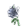
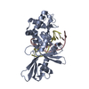
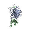
| Title | Crystal structure of MCFD2 in complex with carbohydrate recognition domain of ERGIC-53 | ||||||
 Components Components |
| ||||||
 Keywords Keywords | PROTEIN TRANSPORT / LECTIN / ERGIC / ER / GOLGI / TRANSPORT / Disulfide bond / Endoplasmic reticulum / ER-Golgi transport / Golgi apparatus / Membrane / Transmembrane / Disease mutation | ||||||
| Function / homology |  Function and homology information Function and homology informationTransport to the Golgi and subsequent modification / positive regulation of organelle organization / : / Cargo concentration in the ER / COPII-coated ER to Golgi transport vesicle / endoplasmic reticulum organization / RHOD GTPase cycle / COPII-mediated vesicle transport / RHOC GTPase cycle / D-mannose binding ...Transport to the Golgi and subsequent modification / positive regulation of organelle organization / : / Cargo concentration in the ER / COPII-coated ER to Golgi transport vesicle / endoplasmic reticulum organization / RHOD GTPase cycle / COPII-mediated vesicle transport / RHOC GTPase cycle / D-mannose binding / endoplasmic reticulum-Golgi intermediate compartment / Golgi organization / RHOG GTPase cycle / RAC3 GTPase cycle / RHOA GTPase cycle / RAC2 GTPase cycle / endoplasmic reticulum to Golgi vesicle-mediated transport / vesicle-mediated transport / endoplasmic reticulum-Golgi intermediate compartment membrane / sarcomere / ER to Golgi transport vesicle membrane / blood coagulation / : / protein transport / protein folding / extracellular matrix / gene expression / in utero embryonic development / Golgi membrane / calcium ion binding / endoplasmic reticulum membrane / Golgi apparatus / endoplasmic reticulum / extracellular exosome / membrane / metal ion binding Similarity search - Function | ||||||
| Biological species |  Homo sapiens (human) Homo sapiens (human) | ||||||
| Method |  X-RAY DIFFRACTION / SYNCHROTRON / MOLECULAR REPLACEMENT / Resolution: 1.84 Å X-RAY DIFFRACTION / SYNCHROTRON / MOLECULAR REPLACEMENT / Resolution: 1.84 Å | ||||||
 Authors Authors | Nishio, M. / Kamiya, Y. / Mizushima, T. / Wakatsuki, S. / Sasakawa, H. / Yamamoto, K. / Uchiyama, S. / Noda, M. / McKay, A.R. / Fukui, K. ...Nishio, M. / Kamiya, Y. / Mizushima, T. / Wakatsuki, S. / Sasakawa, H. / Yamamoto, K. / Uchiyama, S. / Noda, M. / McKay, A.R. / Fukui, K. / Hauri, H.P. / Kato, K. | ||||||
 Citation Citation | Journal: Proc.Natl.Acad.Sci.USA / Year: 2010 Title: Structural basis for the cooperative interplay between the two causative gene products of combined factor V and factor VIII deficiency. Authors: Nishio, M. / Kamiya, Y. / Mizushima, T. / Wakatsuki, S. / Sasakawa, H. / Yamamoto, K. / Uchiyama, S. / Noda, M. / McKay, A.R. / Fukui, K. / Hauri, H.P. / Kato, K. | ||||||
| History |
|
- Structure visualization
| Structure viewer | Molecule: MolmilJmol/JSmol |
|---|
- Downloads & links
-Download
| PDBx/mmCIF format | 3a4u.cif.gz | 81 KB | Display | PDBx/mmCIF format |
|---|---|---|---|---|
| PDB format | pdb3a4u.ent.gz | 58.3 KB | Display | PDB format |
| PDBx/mmJSON format | 3a4u.json.gz | Tree view | PDBx/mmJSON format | |
| Others |  Other downloads Other downloads |
-Validation report
| Arichive directory | https://data.pdbj.org/pub/pdb/validation_reports/a4/3a4uftp://data.pdbj.org/pub/pdb/validation_reports/a4/3a4u | HTTPS FTP |
|---|
-Related structure data
| Related structure data | 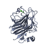 1r1zS S: Starting model for refinement |
|---|---|
| Similar structure data |


-Links
 PDBj
PDBj


- Assembly
| Deposited unit | 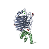
| ||||||||
|---|---|---|---|---|---|---|---|---|---|
| 1 |
| ||||||||
| Unit cell |
|
-Components
| #1: Protein | Mass: 28039.068 Da / Num. of mol.: 1 / Fragment: Carbohydrate Recognition Domain Source method: isolated from a genetically manipulated source Source: (gene. exp.) Homo sapiens (human) / Gene: LMAN1, ERGIC53, F5F8D / Plasmid: pCold3 / Production host:  | ||||||
|---|---|---|---|---|---|---|---|
| #2: Protein | Mass: 16316.842 Da / Num. of mol.: 1 Source method: isolated from a genetically manipulated source Source: (gene. exp.) Homo sapiens (human) / Gene: MCFD2, SDNSF / Plasmid: pET16b / Production host: | ||||||
| #3: Chemical | ChemComp-CA /   Mass: 40.078 Da / Num. of mol.: 4 / Source method: obtained synthetically / Formula: Ca Mass: 40.078 Da / Num. of mol.: 4 / Source method: obtained synthetically / Formula: Ca#4: Chemical | ChemComp-GOL /   Mass: 92.094 Da / Num. of mol.: 4 / Source method: obtained synthetically / Formula: C3H8O3 Mass: 92.094 Da / Num. of mol.: 4 / Source method: obtained synthetically / Formula: C3H8O3#5: Water | ChemComp-HOH / |  Mass: 18.015 Da / Num. of mol.: 189 / Source method: isolated from a natural source / Formula: H2O Mass: 18.015 Da / Num. of mol.: 189 / Source method: isolated from a natural source / Formula: H2OHas protein modification | Y | |
-Experimental details
-Experiment
| Experiment | Method: X-RAY DIFFRACTION / Number of used crystals: 1 |
|---|
- Sample preparation
| Crystal | Density Matthews: 2.28 Å3/Da / Density % sol: 46.14 % |
|---|---|
| Crystal grow | Temperature: 289 K / Method: vapor diffusion, hanging drop / pH: 5.5 Details: 20% PEG 3350, 0.2M sodium tartrate, pH 5.5, VAPOR DIFFUSION, HANGING DROP, temperature 289K |
-Data collection
| Diffraction | Mean temperature: 100 K |
|---|---|
| Diffraction source | Source: SYNCHROTRON / Site: Photon Factory  / Beamline: BL-5A / Wavelength: 1 Å / Beamline: BL-5A / Wavelength: 1 Å |
| Detector | Type: ADSC QUANTUM 315r / Detector: CCD / Date: Dec 13, 2008 / Details: mirrors |
| Radiation | Protocol: SINGLE WAVELENGTH / Monochromatic (M) / Laue (L): M / Scattering type: x-ray |
| Radiation wavelength | Wavelength: 1 Å / Relative weight: 1 |
| Reflection | Resolution: 1.84→49.81 Å / Num. obs: 36530 / % possible obs: 99.9 % / Redundancy: 6.8 % / Biso Wilson estimate: 19.84 Å2 / Rmerge(I) obs: 0.059 / Rsym value: 0.188 / Net I/σ(I): 19.5 |
| Reflection shell | Resolution: 1.84→1.94 Å / Redundancy: 7 % / Rmerge(I) obs: 0.257 / Mean I/σ(I) obs: 6.1 / Num. unique all: 5229 / Rsym value: 0.188 / % possible all: 100 |
- Processing
| Software |
| ||||||||||||||||||||||||||||||||||||||||||||||||||||||||||||||||||||||||||||||||||||||||||||||||||||||||||||||||||||||||||||||||||||||||||||||||||||||||||||||||||||||||||
|---|---|---|---|---|---|---|---|---|---|---|---|---|---|---|---|---|---|---|---|---|---|---|---|---|---|---|---|---|---|---|---|---|---|---|---|---|---|---|---|---|---|---|---|---|---|---|---|---|---|---|---|---|---|---|---|---|---|---|---|---|---|---|---|---|---|---|---|---|---|---|---|---|---|---|---|---|---|---|---|---|---|---|---|---|---|---|---|---|---|---|---|---|---|---|---|---|---|---|---|---|---|---|---|---|---|---|---|---|---|---|---|---|---|---|---|---|---|---|---|---|---|---|---|---|---|---|---|---|---|---|---|---|---|---|---|---|---|---|---|---|---|---|---|---|---|---|---|---|---|---|---|---|---|---|---|---|---|---|---|---|---|---|---|---|---|---|---|---|---|---|---|
| Refinement | Method to determine structure: MOLECULAR REPLACEMENT Starting model: PDB ENTRY 1R1Z Resolution: 1.84→49.81 Å / Cor.coef. Fo:Fc: 0.951 / Cor.coef. Fo:Fc free: 0.944 / SU B: 1.975 / SU ML: 0.062 / Cross valid method: THROUGHOUT / ESU R: 0.113 / ESU R Free: 0.101 / Stereochemistry target values: MAXIMUM LIKELIHOOD / Details: HYDROGENS HAVE BEEN ADDED IN THE RIDING POSITIONS
| ||||||||||||||||||||||||||||||||||||||||||||||||||||||||||||||||||||||||||||||||||||||||||||||||||||||||||||||||||||||||||||||||||||||||||||||||||||||||||||||||||||||||||
| Solvent computation | Ion probe radii: 0.8 Å / Shrinkage radii: 0.8 Å / VDW probe radii: 1.4 Å / Solvent model: MASK | ||||||||||||||||||||||||||||||||||||||||||||||||||||||||||||||||||||||||||||||||||||||||||||||||||||||||||||||||||||||||||||||||||||||||||||||||||||||||||||||||||||||||||
| Displacement parameters | Biso mean: 21.197 Å2
| ||||||||||||||||||||||||||||||||||||||||||||||||||||||||||||||||||||||||||||||||||||||||||||||||||||||||||||||||||||||||||||||||||||||||||||||||||||||||||||||||||||||||||
| Refinement step | Cycle: LAST / Resolution: 1.84→49.81 Å
| ||||||||||||||||||||||||||||||||||||||||||||||||||||||||||||||||||||||||||||||||||||||||||||||||||||||||||||||||||||||||||||||||||||||||||||||||||||||||||||||||||||||||||
| Refine LS restraints |
| ||||||||||||||||||||||||||||||||||||||||||||||||||||||||||||||||||||||||||||||||||||||||||||||||||||||||||||||||||||||||||||||||||||||||||||||||||||||||||||||||||||||||||
| LS refinement shell | Resolution: 1.838→1.886 Å / Total num. of bins used: 20
|
