 Movie
Movie Controller

[English] 日本語
 Yorodumi
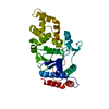
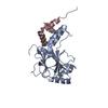
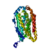
- PDB-3idq: Crystal structure of S. cerevisiae Get3 at 3.7 Angstrom resolution -
+ Open data

- Basic information
| Entry | Database: PDB / ID: 3idq | ||||||
|---|---|---|---|---|---|---|---|
| Title | Crystal structure of S. cerevisiae Get3 at 3.7 Angstrom resolution | ||||||
 Components Components | ATPase GET3 | ||||||
 Keywords Keywords | HYDROLASE / deviant Walker A motif / Arsenical resistance / ATP-binding / Cytoplasm / Endoplasmic reticulum / ER-Golgi transport / Golgi apparatus / Nucleotide-binding / Transport | ||||||
| Function / homology |  Function and homology information Function and homology informationGET complex / : / tail-anchored membrane protein insertion into ER membrane / Hydrolases; Acting on acid anhydrides / protein insertion into ER membrane / post-translational protein targeting to endoplasmic reticulum membrane / response to arsenic-containing substance / retrograde vesicle-mediated transport, Golgi to endoplasmic reticulum / response to metal ion / protein folding chaperone ...GET complex / : / tail-anchored membrane protein insertion into ER membrane / Hydrolases; Acting on acid anhydrides / protein insertion into ER membrane / post-translational protein targeting to endoplasmic reticulum membrane / response to arsenic-containing substance / retrograde vesicle-mediated transport, Golgi to endoplasmic reticulum / response to metal ion / protein folding chaperone / guanyl-nucleotide exchange factor activity / : / response to heat / cellular response to oxidative stress / endoplasmic reticulum membrane / Golgi apparatus / endoplasmic reticulum / ATP hydrolysis activity / ATP binding / metal ion binding / identical protein binding / cytosol Similarity search - Function | ||||||
| Biological species |  | ||||||
| Method |  X-RAY DIFFRACTION / SYNCHROTRON / MOLECULAR REPLACEMENT / Resolution: 3.701 Å X-RAY DIFFRACTION / SYNCHROTRON / MOLECULAR REPLACEMENT / Resolution: 3.701 Å | ||||||
 Authors Authors | Suloway, C.J.M. / Chartron, J.W. / Zaslaver, M. / Clemons Jr., W.M. | ||||||
 Citation Citation | Journal: Proc.Natl.Acad.Sci.USA / Year: 2009 Title: Model for eukaryotic tail-anchored protein binding based on the structure of Get3 Authors: Suloway, C.J. / Chartron, J.W. / Zaslaver, M. / Clemons, W.M. | ||||||
| History |
|
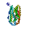
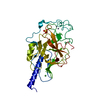
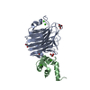
- Structure visualization
| Structure viewer | Molecule: MolmilJmol/JSmol |
|---|
- Downloads & links
-Download
| PDBx/mmCIF format | 3idq.cif.gz | 64.3 KB | Display | PDBx/mmCIF format |
|---|---|---|---|---|
| PDB format | pdb3idq.ent.gz | 46.9 KB | Display | PDB format |
| PDBx/mmJSON format | 3idq.json.gz | Tree view | PDBx/mmJSON format | |
| Others |  Other downloads Other downloads |
-Validation report
| Arichive directory | https://data.pdbj.org/pub/pdb/validation_reports/id/3idqftp://data.pdbj.org/pub/pdb/validation_reports/id/3idq | HTTPS FTP |
|---|
-Related structure data
| Related structure data |  3ibgC 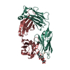 1ibgS S: Starting model for refinement C: citing same article ( |
|---|---|
| Similar structure data |


-Links
 PDBj
PDBj- Assembly
| Deposited unit | 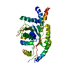
| ||||||||
|---|---|---|---|---|---|---|---|---|---|
| 1 |
| ||||||||
| 2 | 
| ||||||||
| Unit cell |
| ||||||||
| Components on special symmetry positions |
|
-Components
| #1: Protein | Mass: 41132.508 Da / Num. of mol.: 1 Source method: isolated from a genetically manipulated source Source: (gene. exp.) Gene: GET3, ARR4, YDL100C, D2371 / Plasmid: pET33b / Production host:  |
|---|---|
| #2: Chemical | ChemComp-NI /   Mass: 58.693 Da / Num. of mol.: 1 / Source method: obtained synthetically / Formula: Ni Mass: 58.693 Da / Num. of mol.: 1 / Source method: obtained synthetically / Formula: Ni |
| #3: Chemical | ChemComp-ZN /   Mass: 65.409 Da / Num. of mol.: 1 / Source method: obtained synthetically / Formula: Zn Mass: 65.409 Da / Num. of mol.: 1 / Source method: obtained synthetically / Formula: Zn |
-Experimental details
-Experiment
| Experiment | Method: X-RAY DIFFRACTION / Number of used crystals: 1 |
|---|
- Sample preparation
| Crystal | Density Matthews: 4.37 Å3/Da / Density % sol: 71.87 % |
|---|---|
| Crystal grow | Temperature: 298 K / Method: vapor diffusion, sitting drop / pH: 8 Details: 0.1 M HEPES, 1.6 M ammonium sulfate, pH 8.0, VAPOR DIFFUSION, SITTING DROP, temperature 298K |
-Data collection
| Diffraction | Mean temperature: 100 K |
|---|---|
| Diffraction source | Source: SYNCHROTRON / Site: SSRL  / Beamline: BL12-2 / Wavelength: 1 Å / Beamline: BL12-2 / Wavelength: 1 Å |
| Detector | Type: MARMOSAIC 325 mm CCD / Detector: CCD / Date: May 15, 2009 |
| Radiation | Monochromator: LIQUID NITROGEN-COOLED DOUBLE CRYSTAL / Protocol: SINGLE WAVELENGTH / Monochromatic (M) / Laue (L): M / Scattering type: x-ray |
| Radiation wavelength | Wavelength: 1 Å / Relative weight: 1 |
| Reflection | Resolution: 3.701→47.054 Å / Num. obs: 7954 / % possible obs: 99.9 % / Redundancy: 5.9 % / Rsym value: 0.099 |
| Reflection shell | Resolution: 3.7→3.9 Å / Redundancy: 6 % / Rsym value: 0.627 / % possible all: 100 |
- Processing
| Software |
| ||||||||||||||||||||||||||||||||
|---|---|---|---|---|---|---|---|---|---|---|---|---|---|---|---|---|---|---|---|---|---|---|---|---|---|---|---|---|---|---|---|---|---|
| Refinement | Method to determine structure: MOLECULAR REPLACEMENT Starting model: PDB entry 1IBG Resolution: 3.701→47.054 Å / Occupancy max: 1 / Occupancy min: 0.33 / SU ML: 0.54 / σ(F): 1.34 / Stereochemistry target values: ML Details: Residues designated 365-369 are part of the histidine tag used in purification. Density connecting these residues to the rest of the protein in chain A was not observed, therefore it is ...Details: Residues designated 365-369 are part of the histidine tag used in purification. Density connecting these residues to the rest of the protein in chain A was not observed, therefore it is possible they are extending from a symmetry related copy.
| ||||||||||||||||||||||||||||||||
| Solvent computation | Shrinkage radii: 0.9 Å / VDW probe radii: 1.11 Å / Solvent model: FLAT BULK SOLVENT MODEL / Bsol: 150 Å2 / ksol: 0.32 e/Å3 | ||||||||||||||||||||||||||||||||
| Displacement parameters | Biso max: 401.21 Å2 / Biso mean: 182.045 Å2 / Biso min: 59 Å2
| ||||||||||||||||||||||||||||||||
| Refinement step | Cycle: LAST / Resolution: 3.701→47.054 Å
| ||||||||||||||||||||||||||||||||
| Refine LS restraints |
| ||||||||||||||||||||||||||||||||
| LS refinement shell | Refine-ID: X-RAY DIFFRACTION / Total num. of bins used: 3
|
