 Movie
Movie Controller


+ Open data

- Basic information
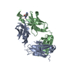
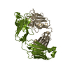
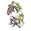
| Entry | Database: PDB / ID: 1ibg | ||||||
|---|---|---|---|---|---|---|---|
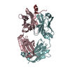
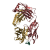
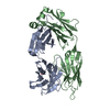
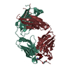
| Title | STRUCTURE AND SPECIFICITY OF THE ANTI-DIGOXIN ANTIBODY 40-50 | ||||||
 Components Components |
| ||||||
 Keywords Keywords | IMMUNE SYSTEM / IMMUNOGLOBULIN / FAB / COMPLEX | ||||||
| Function / homology | Immunoglobulins / Immunoglobulin-like / Sandwich / Mainly Beta / COPPER (II) ION / OUABAIN / :  Function and homology information Function and homology information | ||||||
| Biological species |  | ||||||
| Method |  X-RAY DIFFRACTION / Resolution: 2.7 Å X-RAY DIFFRACTION / Resolution: 2.7 Å | ||||||
 Authors Authors | Jeffrey, P.D. / Sheriff, S. | ||||||
 Citation Citation | Journal: J.Mol.Biol. / Year: 1995 Title: Structure and Specificity of the Anti-Digoxin Antibody 40-50 Authors: Jeffrey, P.D. / Schildbach, J.F. / Chang, C.Y. / Kussie, P.H. / Margolies, M.N. / Sheriff, S. #1: Journal: To be PublishedTitle: Crystallization and Preliminary X-Ray Analysis of Anti-Digoxin Antibodies Authors: Chang, C.Y. / Shih, H. / Jeffrey, P.D. / Margolies, M.N. / Sheriff, S. | ||||||
| History |
|
- Structure visualization
| Structure viewer | Molecule: MolmilJmol/JSmol |
|---|
- Downloads & links
-Download
| PDBx/mmCIF format | 1ibg.cif.gz | 98.6 KB | Display | PDBx/mmCIF format |
|---|---|---|---|---|
| PDB format | pdb1ibg.ent.gz | 73.3 KB | Display | PDB format |
| PDBx/mmJSON format | 1ibg.json.gz | Tree view | PDBx/mmJSON format | |
| Others |  Other downloads Other downloads |
-Validation report
| Arichive directory | https://data.pdbj.org/pub/pdb/validation_reports/ib/1ibgftp://data.pdbj.org/pub/pdb/validation_reports/ib/1ibg | HTTPS FTP |
|---|
-Related structure data
| Similar structure data |
|---|


-Links
 PDBj
PDBj


- Assembly
| Deposited unit | 
| ||||||||
|---|---|---|---|---|---|---|---|---|---|
| 1 |
| ||||||||
| 2 | 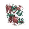
| ||||||||
| Unit cell |
| ||||||||
| Atom site foot note | 1: CIS PROLINE - PRO L 8 / 2: CIS PROLINE - PRO L 77 / 3: CIS PROLINE - PRO L 95 / 4: CIS PROLINE - PRO L 141 5: GLY H 8 - PRO H 9 OMEGA = 236.30 PEPTIDE BOND DEVIATES SIGNIFICANTLY FROM TRANS CONFORMATION 6: CIS PROLINE - PRO H 149 / 7: CIS PROLINE - PRO H 151 8: TRP H 199 - PRO H 200 OMEGA = 215.31 PEPTIDE BOND DEVIATES SIGNIFICANTLY FROM TRANS CONFORMATION |
-Components
| #1: Antibody | Mass: 23842.357 Da / Num. of mol.: 1 Source method: isolated from a genetically manipulated source Source: (gene. exp.) |
|---|---|
| #2: Antibody | Mass: 23146.904 Da / Num. of mol.: 1 Source method: isolated from a genetically manipulated source Source: (gene. exp.) |
| #3: Chemical | ChemComp-CU /   Mass: 63.546 Da / Num. of mol.: 1 / Source method: obtained synthetically / Formula: Cu Mass: 63.546 Da / Num. of mol.: 1 / Source method: obtained synthetically / Formula: Cu |
| #4: Chemical | ChemComp-OBN /   Mass: 584.652 Da / Num. of mol.: 1 / Source method: obtained synthetically / Formula: C29H44O12 Mass: 584.652 Da / Num. of mol.: 1 / Source method: obtained synthetically / Formula: C29H44O12 |
| #5: Water | ChemComp-HOH /  Mass: 18.015 Da / Num. of mol.: 1 / Source method: isolated from a natural source / Formula: H2O Mass: 18.015 Da / Num. of mol.: 1 / Source method: isolated from a natural source / Formula: H2O |
| Has protein modification | Y |
| Sequence details | THE FAB LIGHT CHAIN (RESIDUES 1 - 214) HAS BEEN ASSIGNED CHAIN INDICATOR L. THE FAB HEAVY CHAIN ...THE FAB LIGHT CHAIN (RESIDUES 1 - 214) HAS BEEN ASSIGNED CHAIN INDICATOR L. THE FAB HEAVY CHAIN (RESIDUES 1 - 223) HAS BEEN ASSIGNED CHAIN INDICATOR H. THE FAB FRAGMENT IS NUMBERED BY THE CONVENTION |
-Experimental details
-Experiment
| Experiment | Method: X-RAY DIFFRACTION |
|---|
- Sample preparation
| Crystal | Density Matthews: 2.33 Å3/Da / Density % sol: 47.28 % | ||||||||||||||||||||||||||||||||||||||||||||||||
|---|---|---|---|---|---|---|---|---|---|---|---|---|---|---|---|---|---|---|---|---|---|---|---|---|---|---|---|---|---|---|---|---|---|---|---|---|---|---|---|---|---|---|---|---|---|---|---|---|---|
| Crystal | *PLUS Density % sol: 44 % | ||||||||||||||||||||||||||||||||||||||||||||||||
| Crystal grow | *PLUS pH: 8 / Method: vapor diffusion | ||||||||||||||||||||||||||||||||||||||||||||||||
| Components of the solutions | *PLUS
|
-Data collection
| Radiation | Scattering type: x-ray |
|---|---|
| Radiation wavelength | Relative weight: 1 |
| Reflection | Num. obs: 12688 |
| Reflection | *PLUS Highest resolution: 2.43 Å / Num. measured all: 24232 / Rmerge(I) obs: 0.049 |
- Processing
| Software |
| ||||||||||||||||||||||||||||||||||||||||||||||||||||||||||||
|---|---|---|---|---|---|---|---|---|---|---|---|---|---|---|---|---|---|---|---|---|---|---|---|---|---|---|---|---|---|---|---|---|---|---|---|---|---|---|---|---|---|---|---|---|---|---|---|---|---|---|---|---|---|---|---|---|---|---|---|---|---|
| Refinement | Resolution: 2.7→10 Å / σ(F): 1 /
| ||||||||||||||||||||||||||||||||||||||||||||||||||||||||||||
| Displacement parameters | Biso mean: 31.4 Å2 | ||||||||||||||||||||||||||||||||||||||||||||||||||||||||||||
| Refine analyze | Luzzati coordinate error obs: 0.35 Å | ||||||||||||||||||||||||||||||||||||||||||||||||||||||||||||
| Refinement step | Cycle: LAST / Resolution: 2.7→10 Å
| ||||||||||||||||||||||||||||||||||||||||||||||||||||||||||||
| Refine LS restraints |
| ||||||||||||||||||||||||||||||||||||||||||||||||||||||||||||
| Refinement | *PLUS | ||||||||||||||||||||||||||||||||||||||||||||||||||||||||||||
| Solvent computation | *PLUS | ||||||||||||||||||||||||||||||||||||||||||||||||||||||||||||
| Displacement parameters | *PLUS | ||||||||||||||||||||||||||||||||||||||||||||||||||||||||||||
| Refine LS restraints | *PLUS
|
