 Movie
Movie Controller

[English] 日本語
 Yorodumi
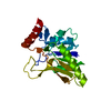
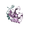
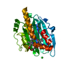
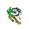
- PDB-1r1z: The Crystal structure of the Carbohydrate recognition domain of t... -
+ Open data

- Basic information
| Entry | Database: PDB / ID: 1r1z | ||||||
|---|---|---|---|---|---|---|---|
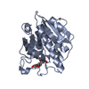
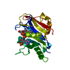
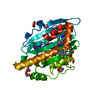
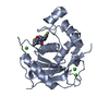
| Title | The Crystal structure of the Carbohydrate recognition domain of the glycoprotein sorting receptor p58/ERGIC-53 reveals a novel metal binding site and conformational changes associated with calcium ion binding | ||||||
 Components Components | ERGIC-53 protein | ||||||
 Keywords Keywords | SUGAR BINDING PROTEIN / beta-sheet / calcium-binding / lectin / mammalian / endoplasmic reticulum | ||||||
| Function / homology |  Function and homology information Function and homology informationcargo receptor complex / RAC3 GTPase cycle / RHOC GTPase cycle / postsynaptic Golgi apparatus / RAC2 GTPase cycle / RHOG GTPase cycle / RHOA GTPase cycle / Cargo concentration in the ER / COPII-mediated vesicle transport / COPII-coated ER to Golgi transport vesicle ...cargo receptor complex / RAC3 GTPase cycle / RHOC GTPase cycle / postsynaptic Golgi apparatus / RAC2 GTPase cycle / RHOG GTPase cycle / RHOA GTPase cycle / Cargo concentration in the ER / COPII-mediated vesicle transport / COPII-coated ER to Golgi transport vesicle / endoplasmic reticulum organization / D-mannose binding / endoplasmic reticulum-Golgi intermediate compartment / Golgi organization / endoplasmic reticulum to Golgi vesicle-mediated transport / endoplasmic reticulum-Golgi intermediate compartment membrane / sarcomere / protein transport / gene expression / in utero embryonic development / Golgi membrane / calcium ion binding / endoplasmic reticulum membrane / glutamatergic synapse / Golgi apparatus / endoplasmic reticulum / metal ion binding / identical protein binding Similarity search - Function | ||||||
| Biological species |  | ||||||
| Method |  X-RAY DIFFRACTION / SYNCHROTRON / MOLECULAR REPLACEMENT / Resolution: 2.4 Å X-RAY DIFFRACTION / SYNCHROTRON / MOLECULAR REPLACEMENT / Resolution: 2.4 Å | ||||||
 Authors Authors | Velloso, L.M. / Svensson, K. / Pettersson, R.F. / Lindqvist, Y. | ||||||
 Citation Citation | Journal: J.Mol.Biol. / Year: 2003 Title: The Crystal Structure of the Carbohydrate-recognition Domain of the Glycoprotein Sorting Receptor p58/ERGIC-53 Reveals an Unpredicted Metal-binding Site and Conformational Changes Associated ...Title: The Crystal Structure of the Carbohydrate-recognition Domain of the Glycoprotein Sorting Receptor p58/ERGIC-53 Reveals an Unpredicted Metal-binding Site and Conformational Changes Associated with Calcium Ion Binding. Authors: Velloso, L.M. / Svensson, K. / Pettersson, R.F. / Lindqvist, Y. | ||||||
| History |
|
- Structure visualization
| Structure viewer | Molecule: MolmilJmol/JSmol |
|---|
- Downloads & links
-Download
| PDBx/mmCIF format | 1r1z.cif.gz | 203.7 KB | Display | PDBx/mmCIF format |
|---|---|---|---|---|
| PDB format | pdb1r1z.ent.gz | 160.8 KB | Display | PDB format |
| PDBx/mmJSON format | 1r1z.json.gz | Tree view | PDBx/mmJSON format | |
| Others |  Other downloads Other downloads |
-Validation report
| Arichive directory | https://data.pdbj.org/pub/pdb/validation_reports/r1/1r1zftp://data.pdbj.org/pub/pdb/validation_reports/r1/1r1z | HTTPS FTP |
|---|
-Related structure data
| Related structure data | 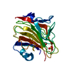 1gv9S S: Starting model for refinement |
|---|---|
| Similar structure data |


-Links
 PDBj
PDBj- Assembly
| Deposited unit | 
| |||||||||||||||||||||||||||||||||||||||||||||||||||||||||||||||||||||||||||||||||||||||||||||||||||||||||||||||||||||
|---|---|---|---|---|---|---|---|---|---|---|---|---|---|---|---|---|---|---|---|---|---|---|---|---|---|---|---|---|---|---|---|---|---|---|---|---|---|---|---|---|---|---|---|---|---|---|---|---|---|---|---|---|---|---|---|---|---|---|---|---|---|---|---|---|---|---|---|---|---|---|---|---|---|---|---|---|---|---|---|---|---|---|---|---|---|---|---|---|---|---|---|---|---|---|---|---|---|---|---|---|---|---|---|---|---|---|---|---|---|---|---|---|---|---|---|---|---|---|
| 1 | 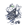
| |||||||||||||||||||||||||||||||||||||||||||||||||||||||||||||||||||||||||||||||||||||||||||||||||||||||||||||||||||||
| 2 | 
| |||||||||||||||||||||||||||||||||||||||||||||||||||||||||||||||||||||||||||||||||||||||||||||||||||||||||||||||||||||
| 3 | 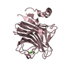
| |||||||||||||||||||||||||||||||||||||||||||||||||||||||||||||||||||||||||||||||||||||||||||||||||||||||||||||||||||||
| 4 | 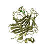
| |||||||||||||||||||||||||||||||||||||||||||||||||||||||||||||||||||||||||||||||||||||||||||||||||||||||||||||||||||||
| Unit cell |
| |||||||||||||||||||||||||||||||||||||||||||||||||||||||||||||||||||||||||||||||||||||||||||||||||||||||||||||||||||||
| Noncrystallographic symmetry (NCS) | NCS domain:
NCS domain segments: Ens-ID: 1 / Refine code: 1
|
-Components
| #1: Protein | Mass: 29027.236 Da / Num. of mol.: 4 Source method: isolated from a genetically manipulated source Source: (gene. exp.)   Spodoptera frugiperda (fall armyworm) / References: UniProt: Q62902 Spodoptera frugiperda (fall armyworm) / References: UniProt: Q62902#2: Chemical | ChemComp-CA /   Mass: 40.078 Da / Num. of mol.: 8 / Source method: obtained synthetically / Formula: Ca Mass: 40.078 Da / Num. of mol.: 8 / Source method: obtained synthetically / Formula: Ca#3: Water | ChemComp-HOH / |  Mass: 18.015 Da / Num. of mol.: 193 / Source method: isolated from a natural source / Formula: H2O Mass: 18.015 Da / Num. of mol.: 193 / Source method: isolated from a natural source / Formula: H2OHas protein modification | Y | |
|---|
-Experimental details
-Experiment
| Experiment | Method: X-RAY DIFFRACTION / Number of used crystals: 1 |
|---|
- Sample preparation
| Crystal | Density Matthews: 2.53 Å3/Da / Density % sol: 51.39 % | ||||||||||||||||||||||||||||||||||||||||||
|---|---|---|---|---|---|---|---|---|---|---|---|---|---|---|---|---|---|---|---|---|---|---|---|---|---|---|---|---|---|---|---|---|---|---|---|---|---|---|---|---|---|---|---|
| Crystal grow | Temperature: 293 K / Method: vapor diffusion, hanging drop / pH: 7.5 Details: PEG8000, Calcium Chloride, Tris, pH 7.5, VAPOR DIFFUSION, HANGING DROP, temperature 293K | ||||||||||||||||||||||||||||||||||||||||||
| Crystal grow | *PLUS Method: vapor diffusion, hanging drop | ||||||||||||||||||||||||||||||||||||||||||
| Components of the solutions | *PLUS
|
-Data collection
| Diffraction | Mean temperature: 100 K |
|---|---|
| Diffraction source | Source: SYNCHROTRON / Site: ESRF  / Beamline: ID14-1 / Wavelength: 0.934 Å / Beamline: ID14-1 / Wavelength: 0.934 Å |
| Detector | Type: ADSC QUANTUM 4 / Detector: CCD / Date: Jul 1, 2002 |
| Radiation | Protocol: SINGLE WAVELENGTH / Monochromatic (M) / Laue (L): M / Scattering type: x-ray |
| Radiation wavelength | Wavelength: 0.934 Å / Relative weight: 1 |
| Reflection | Resolution: 2.4→10 Å / Num. all: 98470 / Num. obs: 42617 / % possible obs: 95.7 % / Observed criterion σ(F): 1 / Observed criterion σ(I): 1 / Redundancy: 2.3 % / Biso Wilson estimate: 45.6 Å2 / Rmerge(I) obs: 0.126 / Rsym value: 0.096 / Net I/σ(I): 13 |
| Reflection shell | Resolution: 2.4→2.53 Å / Redundancy: 2.3 % / Rmerge(I) obs: 0.43 / Mean I/σ(I) obs: 1.7 / Num. unique all: 6205 / Rsym value: 0.33 / % possible all: 95.7 |
| Reflection | *PLUS Lowest resolution: 10 Å / Num. obs: 42111 / % possible obs: 94.6 % / Rmerge(I) obs: 0.092 |
| Reflection shell | *PLUS % possible obs: 95.3 % / Rmerge(I) obs: 0.334 / Mean I/σ(I) obs: 4.2 |
- Processing
| Software |
| ||||||||||||||||||||||||||||||||||||||||||||||||||||||||||||||||||||||
|---|---|---|---|---|---|---|---|---|---|---|---|---|---|---|---|---|---|---|---|---|---|---|---|---|---|---|---|---|---|---|---|---|---|---|---|---|---|---|---|---|---|---|---|---|---|---|---|---|---|---|---|---|---|---|---|---|---|---|---|---|---|---|---|---|---|---|---|---|---|---|---|
| Refinement | Method to determine structure: MOLECULAR REPLACEMENT Starting model: 1gv9 Resolution: 2.4→10 Å / Cor.coef. Fo:Fc: 0.925 / Cor.coef. Fo:Fc free: 0.918 / SU B: 8.516 / SU ML: 0.202 / Cross valid method: THROUGHOUT / σ(F): 0.005 / σ(I): 0.005 / ESU R: 0.49 / ESU R Free: 0.259 / Details: HYDROGENS HAVE BEEN ADDED IN THE RIDING POSITIONS
| ||||||||||||||||||||||||||||||||||||||||||||||||||||||||||||||||||||||
| Solvent computation | Ion probe radii: 0.8 Å / Shrinkage radii: 0.8 Å / VDW probe radii: 1.4 Å / Solvent model: BABINET MODEL WITH MASK | ||||||||||||||||||||||||||||||||||||||||||||||||||||||||||||||||||||||
| Displacement parameters | Biso mean: 21.012 Å2
| ||||||||||||||||||||||||||||||||||||||||||||||||||||||||||||||||||||||
| Refinement step | Cycle: LAST / Resolution: 2.4→10 Å
| ||||||||||||||||||||||||||||||||||||||||||||||||||||||||||||||||||||||
| Refine LS restraints |
| ||||||||||||||||||||||||||||||||||||||||||||||||||||||||||||||||||||||
| Refine LS restraints NCS | Ens-ID: 1 / Number: 1833 / Refine-ID: X-RAY DIFFRACTION
| ||||||||||||||||||||||||||||||||||||||||||||||||||||||||||||||||||||||
| LS refinement shell | Resolution: 2.4→2.462 Å / Total num. of bins used: 20
| ||||||||||||||||||||||||||||||||||||||||||||||||||||||||||||||||||||||
| Refinement | *PLUS Rfactor Rfree: 0.241 / Rfactor Rwork: 0.223 | ||||||||||||||||||||||||||||||||||||||||||||||||||||||||||||||||||||||
| Solvent computation | *PLUS | ||||||||||||||||||||||||||||||||||||||||||||||||||||||||||||||||||||||
| Displacement parameters | *PLUS | ||||||||||||||||||||||||||||||||||||||||||||||||||||||||||||||||||||||
| Refine LS restraints | *PLUS
|
