 Movie
Movie Controller

[English] 日本語
 Yorodumi
- PDB-4ayj: Molecular structure of a metal-independent bacterial glycosyltran... -
+ Open data

- Basic information
| Entry | Database: PDB / ID: 4ayj | |||||||||
|---|---|---|---|---|---|---|---|---|---|---|
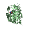
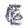
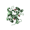
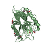
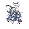
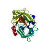
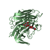
| Title | Molecular structure of a metal-independent bacterial glycosyltransferase that catalyzes the synthesis of histo-blood group A antigen | |||||||||
 Components Components | BOGT - METAL-INDEPENDENT GLYCOSYLTRANSFERASE | |||||||||
 Keywords Keywords | TRANSFERASE / CATALYSIS | |||||||||
| Function / homology |  Function and homology information Function and homology informationhexosyltransferase activity / carbohydrate metabolic process / nucleotide binding / membrane / metal ion binding Similarity search - Function | |||||||||
| Biological species |  BACTEROIDES OVATUS (bacteria) BACTEROIDES OVATUS (bacteria) | |||||||||
| Method |  X-RAY DIFFRACTION / SYNCHROTRON / MOLECULAR REPLACEMENT / Resolution: 3 Å X-RAY DIFFRACTION / SYNCHROTRON / MOLECULAR REPLACEMENT / Resolution: 3 Å | |||||||||
 Authors Authors | Thiyagarajan, N. / Pham, T.T.K. / Stinson, B. / Sundriyal, A. / Tumbale, P. / Lizotte-Waniewskib, M. / Brewb, K. / Acharya, K.R. | |||||||||
 Citation Citation | Journal: Sci.Rep. / Year: 2012 Title: Structure of a Metal-Independent Bacterial Glycosyltransferase that Catalyzes the Synthesis of Histo-Blood Group a Antigen Authors: Thiyagarajan, N. / Pham, T.T.K. / Stinson, B. / Sundriyal, A. / Tumbale, P. / Lizotte-Waniewski, M. / Brew, K. / Acharya, K.R. | |||||||||
| History |
|
- Structure visualization
| Structure viewer | Molecule: MolmilJmol/JSmol |
|---|
- Downloads & links
-Download
| PDBx/mmCIF format | 4ayj.cif.gz | 203 KB | Display | PDBx/mmCIF format |
|---|---|---|---|---|
| PDB format | pdb4ayj.ent.gz | 163.9 KB | Display | PDB format |
| PDBx/mmJSON format | 4ayj.json.gz | Tree view | PDBx/mmJSON format | |
| Others |  Other downloads Other downloads |
-Validation report
| Arichive directory | https://data.pdbj.org/pub/pdb/validation_reports/ay/4ayjftp://data.pdbj.org/pub/pdb/validation_reports/ay/4ayj | HTTPS FTP |
|---|
-Related structure data
| Related structure data | 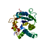 4aylSC S: Starting model for refinement C: citing same article ( |
|---|---|
| Similar structure data |


-Links
 PDBj
PDBj

- Assembly
| Deposited unit | 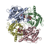
| ||||||||
|---|---|---|---|---|---|---|---|---|---|
| 1 | 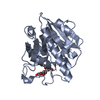
| ||||||||
| 2 | 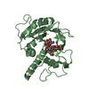
| ||||||||
| 3 | 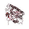
| ||||||||
| 4 | 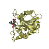
| ||||||||
| Unit cell |
|
-Components
| #1: Protein | Mass: 29038.057 Da / Num. of mol.: 4 / Fragment: RESIDUES 1-246 Source method: isolated from a genetically manipulated source Source: (gene. exp.) BACTEROIDES OVATUS (bacteria) / Production host: References: UniProt: A7LVT2, glycoprotein-fucosylgalactoside alpha-N-acetylgalactosaminyltransferase #2: Polysaccharide | alpha-L-fucopyranose-(1-2)-beta-D-galactopyranose-(1-4)-beta-D-glucopyranose / 2'-fucosyllactose   Source method: isolated from a genetically manipulated source Details: oligosaccharide / References: 2'-fucosyllactose |
|---|
-Experimental details
-Experiment
| Experiment | Method: X-RAY DIFFRACTION / Number of used crystals: 1 |
|---|
- Sample preparation
| Crystal | Density Matthews: 2.16 Å3/Da / Density % sol: 48 % / Description: NONE |
|---|---|
| Crystal grow | Temperature: 289 K / Method: vapor diffusion, sitting drop / pH: 8.5 Details: BOGT6A (4 MG/ML) AND SACCHARIDE (10 MM) WERE INCUBATED OVERNIGHT AT 16 DEG C AND CRYSTALS WERE GROWN IN A WELL SOLUTION CONTAINING 20 % PEG 3350, 0.1 M BIS-TRIS PROPANE, PH 8.5 AND 0.2 M NA/K TARTRATE |
-Data collection
| Diffraction | Mean temperature: 100 K |
|---|---|
| Diffraction source | Source: SYNCHROTRON / Site: Diamond  / Beamline: I02 / Wavelength: 0.9795 / Beamline: I02 / Wavelength: 0.9795 |
| Detector | Type: ADSC QUANTUM 315 / Detector: CCD / Date: Mar 7, 2009 |
| Radiation | Protocol: SINGLE WAVELENGTH / Monochromatic (M) / Laue (L): M / Scattering type: x-ray |
| Radiation wavelength | Wavelength: 0.9795 Å / Relative weight: 1 |
| Reflection | Resolution: 3→70.7 Å / Num. obs: 18669 / % possible obs: 94.1 % / Observed criterion σ(I): 0 / Redundancy: 3.9 % / Biso Wilson estimate: 47.78 Å2 / Rmerge(I) obs: 0.13 / Net I/σ(I): 9.3 |
| Reflection shell | Resolution: 3→3.16 Å / Redundancy: 3.8 % / Rmerge(I) obs: 0.5 / Mean I/σ(I) obs: 2.6 / % possible all: 93.7 |
- Processing
| Software |
| ||||||||||||||||||||||||||||||||||||||||||||||||||||||||
|---|---|---|---|---|---|---|---|---|---|---|---|---|---|---|---|---|---|---|---|---|---|---|---|---|---|---|---|---|---|---|---|---|---|---|---|---|---|---|---|---|---|---|---|---|---|---|---|---|---|---|---|---|---|---|---|---|---|
| Refinement | Method to determine structure: MOLECULAR REPLACEMENT Starting model: PDB ENTRY 4AYL Resolution: 3→70.693 Å / SU ML: 0.92 / σ(F): 1.36 / Phase error: 28.61 / Stereochemistry target values: ML
| ||||||||||||||||||||||||||||||||||||||||||||||||||||||||
| Solvent computation | Shrinkage radii: 0.83 Å / VDW probe radii: 1.1 Å / Solvent model: FLAT BULK SOLVENT MODEL / Bsol: 21.329 Å2 / ksol: 0.307 e/Å3 | ||||||||||||||||||||||||||||||||||||||||||||||||||||||||
| Displacement parameters |
| ||||||||||||||||||||||||||||||||||||||||||||||||||||||||
| Refinement step | Cycle: LAST / Resolution: 3→70.693 Å
| ||||||||||||||||||||||||||||||||||||||||||||||||||||||||
| Refine LS restraints |
| ||||||||||||||||||||||||||||||||||||||||||||||||||||||||
| LS refinement shell |
|
