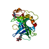
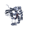
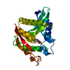
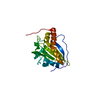
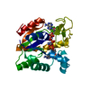
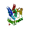
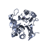
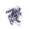
RNAdemethylaseALKBH5 / Alkylated DNA repair protein alkB homolog 5 / Alpha-ketoglutarate-dependent dioxygenase alkB homolog 5
Mass: 25339.010 Da / Num. of mol.: 2 / Fragment: UNP residues 74-294 Source method: isolated from a genetically manipulated source Source: (gene. exp.) Homo sapiens (human) / Gene: ALKBH5, ABH5, OFOXD1 / Plasmid: pET28-MHL / Production host: Escherichia coli (E. coli) / Strain (production host): BL21-V2R-pRARE2 References: UniProt: Q6P6C2, Oxidoreductases; Acting on paired donors, with incorporation or reduction of molecular oxygen; With 2-oxoglutarate as one donor, and incorporation of one atom of oxygen into each donor
Mass: 18.015 Da / Num. of mol.: 23 / Source method: isolated from a natural source / Formula: H2O
Has protein modification
Y
-
Experimental details
-
Experiment
Experiment
Method: X-RAY DIFFRACTION / Number of used crystals: 1
-
Sample preparation
Crystal
Density Matthews: 2.2 Å3/Da / Density % sol: 42.4 %
Crystal grow
Temperature: 291 K / Method: vapor diffusion Details: 0.005 M of each manganous sulfate and 2-oxyglutarate were added to the protein stock solution. Reservoir: 20% PEG-3350, 0.2 M diammonium phosphate, vapor diffusion, temperature 291K
Resolution: 2.28→30 Å / Cor.coef. Fo:Fc: 0.934 / Cor.coef. Fo:Fc free: 0.91 / WRfactor Rfree: 0.2464 / WRfactor Rwork: 0.202 / Occupancy max: 1 / Occupancy min: 0.5 / FOM work R set: 0.686 / SU B: 27.978 / SU ML: 0.305 / SU R Cruickshank DPI: 0.4723 / SU Rfree: 0.2846 / Cross valid method: THROUGHOUT / σ(F): 0 / ESU R: 0.472 / ESU R Free: 0.285 / Stereochemistry target values: MAXIMUM LIKELIHOOD Details: PDB entry 2IUW aided in the interpretation of the manganese cation binding site. Coot was used for interactive model adjustments. Molprobity was used for validation of model geometry.
Rfactor
Num. reflection
% reflection
Selection details
Rfree
0.2841
934
5 %
THIN SHELLS (SFTOOLS)
Rwork
0.2382
-
-
-
obs
0.2405
18790
99.76 %
-
Solvent computation
Ion probe radii: 0.8 Å / Shrinkage radii: 0.8 Å / VDW probe radii: 1.2 Å / Solvent model: MASK
In the structure databanks used in Yorodumi, some data are registered as the other names, "COVID-19 virus" and "2019-nCoV". Here are the details of the virus and the list of structure data.
Jan 31, 2019. EMDB accession codes are about to change! (news from PDBe EMDB page)
EMDB accession codes are about to change! (news from PDBe EMDB page)
The allocation of 4 digits for EMDB accession codes will soon come to an end. Whilst these codes will remain in use, new EMDB accession codes will include an additional digit and will expand incrementally as the available range of codes is exhausted. The current 4-digit format prefixed with “EMD-” (i.e. EMD-XXXX) will advance to a 5-digit format (i.e. EMD-XXXXX), and so on. It is currently estimated that the 4-digit codes will be depleted around Spring 2019, at which point the 5-digit format will come into force.
The EM Navigator/Yorodumi systems omit the EMD- prefix.
Related info.:Q: What is EMD? / ID/Accession-code notation in Yorodumi/EM Navigator
Yorodumi is a browser for structure data from EMDB, PDB, SASBDB, etc.
This page is also the successor to EM Navigator detail page, and also detail information page/front-end page for Omokage search.
The word "yorodu" (or yorozu) is an old Japanese word meaning "ten thousand". "mi" (miru) is to see.
Related info.:EMDB / PDB / SASBDB / Comparison of 3 databanks / Yorodumi Search / Aug 31, 2016. New EM Navigator & Yorodumi / Yorodumi Papers / Jmol/JSmol / Function and homology information / Changes in new EM Navigator and Yorodumi
 Movie
Movie Controller

 Yorodumi
 Open data
 Basic information
 Components
 Keywords
 Function and homology information
 Homo sapiens (human)
 X-RAY DIFFRACTION / direct placement / Resolution: 2.28 Å
  Authors
 Citation
 Structure visualization
 Downloads & links
 Other downloads
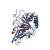


 PDBj
PDBj

 Assembly
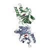


 Mass: 54.938 Da / Num. of mol.: 2 / Source method: obtained synthetically / Formula: Mn
 Mass: 146.098 Da / Num. of mol.: 2 / Source method: obtained synthetically / Formula: C5H6O5
 Num. of mol.: 23 / Source method: obtained synthetically
 Mass: 18.015 Da / Num. of mol.: 23 / Source method: isolated from a natural source / Formula: H2O
 Sample preparation
 Processing